
See-through abdomen display for minimally invasive surgery

a minimally invasive surgery and abdomen technology, applied in the field of minimally invasive surgery, can solve the problems of inconvenient and unclear video orientation, inconvenient operation, and longer completion time than equivalent open surgery

Inactive Publication Date: 2013-08-06
UNIV OF SOUTH FLORIDA
View PDF11 Cites 86 Cited by
  • Summary
  • Abstract
  • Description
  • Claims
  • Application Information

AI Technical Summary

Benefits of technology

The invention is a way to turn images of internal organs and surgical tools into real-life images that can be displayed on a patient's skin. This creates a virtual effect that makes the skin appear transparent, allowing surgeons to see the surgical area without having to look away from the patient. The method uses micro-cameras placed inside the patient's body to capture the images, which are then projected onto the skin. The images are compensated for geometry and color distortion, and can be controlled by the surgeon through gesture recognition and finger tracking. This technology allows for a more intuitive and efficient surgery experience, providing better visual feedback without the risks associated with open-cavity surgery.

Problems solved by technology

While MIS provides many benefits, it often takes longer to complete than equivalent open surgeries.
In particular, MIS is hindered by limited viewpoints and insertion points, inconsistent and unclear orientation of video, and limited touch sensing and hand motion due to long-stick surgical tools.
As a result, MIS requires significantly more training than regular open surgery, which prevents or discourages many surgeons to master the skills for MIS, especially in remote and developing regions or less-than-ideal surgical venues.
The da Vinci® system, however, is very expensive and requires multiple incisions for the robotic arms to perform the operation.
Moreover, the da Vinci® system has unwieldy robotics arms that limit its application; for example, the robotics arms are too big to insert tools near one another and have conflicts with other surgical tools during procedures.
Many surgeons have expressed their disappointment with the fundamental limitations of these scopes based on their experience with hundreds of MIS operations.
Though quite good in image quality, these videoscopes are cumbersome and require a point of access into the patient, either through a separate incision or through a separate trocar site in a multitrocar access port.
The videoscope cables for light, video image, and power clutter and consume space in the operative field.
In addition, the limitation of the viewpoint and view angle of the rigid endoscope is a handicap for surgeons.
The misinterpretation of the image orientation on an overhead monitor also poses a significant problem to the hand-eye coordination for the surgeons and requires great skills and train to master and compensate.
If the mapping does not align correctly with the patient and the surgical tool, the visualization can be dangerous.
It is very challenging to achieve satisfactory accurate alignment between the tracking data and the image since it requires precise models of the patient and models of instruments.

Method used


Image

  • See-through abdomen display for minimally invasive surgery
  • See-through abdomen display for minimally invasive surgery
  • See-through abdomen display for minimally invasive surgery

Examples


Embodiment Construction

[0042]In the following detailed description of the preferred embodiments, reference is made to the accompanying drawings, which form a part hereof, and within which are shown by way of illustration specific embodiments by which the invention may be practiced. It is to be understood that other embodiments may be utilized and structural changes may be made without departing from the scope of the invention

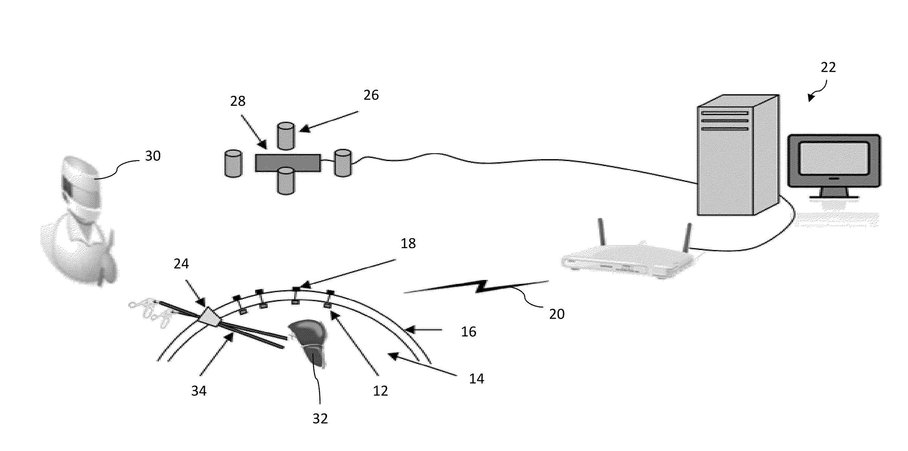
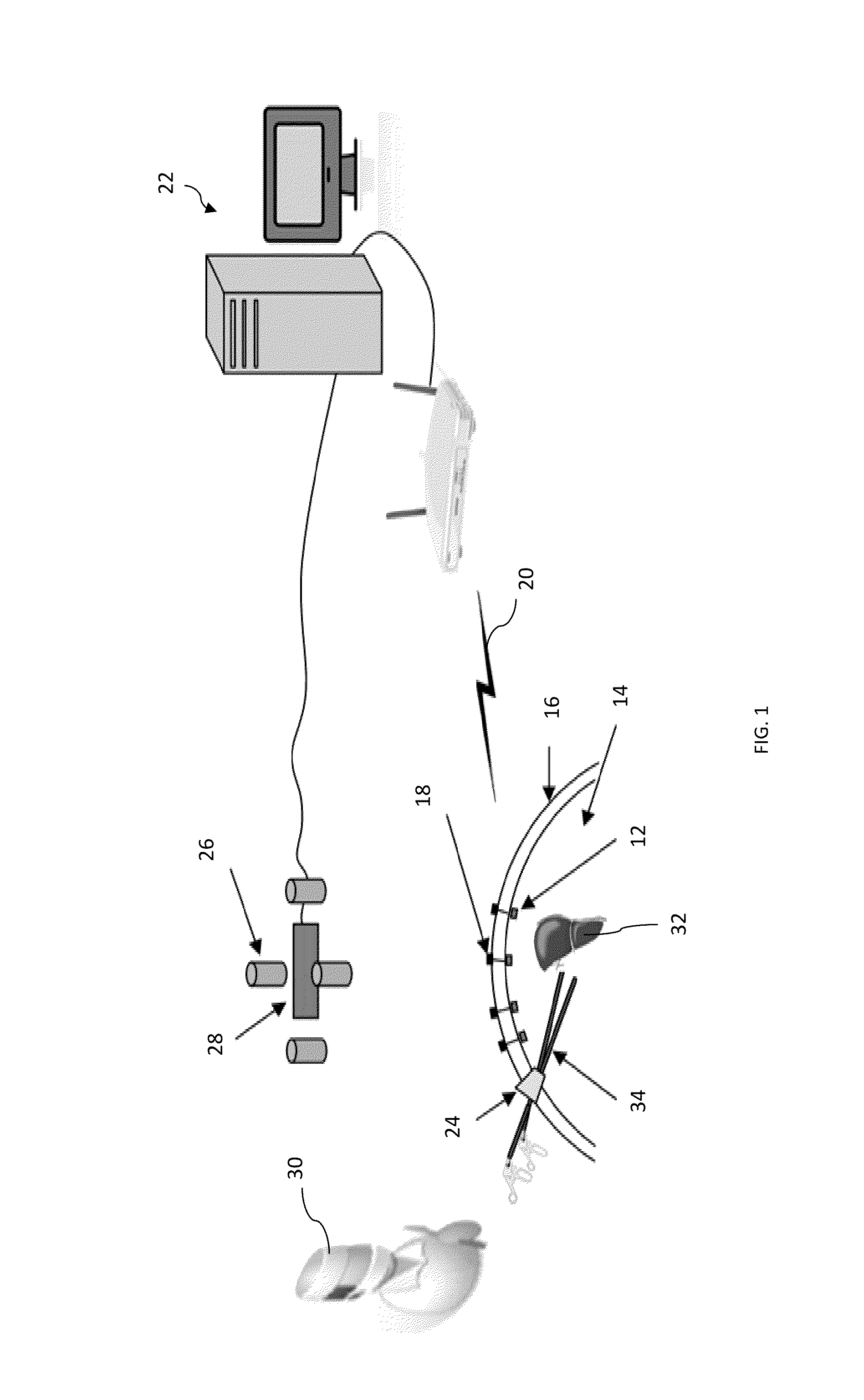
[0043]In an embodiment, as depicted in FIG. 1, the claimed invention includes a plurality of wireless camera modules 12 that are inserted and retreated into a body cavity of interest 14 with a surgery gripper. The tiny wireless camera modules 12 are anchored around the cavity of interest 14 and provide a large view of visual feedback. The feedback is processed in real-time and displayed via projectors 26 on the human body in alignment with the physical cavity of interest 14 and provides a virtually transparent effect so that a surgeon can operate a MIS / LESS procedure with the view of ...


PUM


Abstract

This invention is in the domain of minimally invasive surgery and is a method and apparatus that transforms and displays images of internal organs and tissues taken from internally located imaging devices on external skin. The image displayed on the skin aligns with the actual physical location, orientation, and size of the internal organs and tissues in a way that viewers have the perception that the skin is transparent. This method and apparatus enables surgeons to have the same hand-eye coordination as in an open surgery during a minimally invasive surgery.

Description

CROSS REFERENCE TO RELATED APPLICATIONS[0001]This application claims priority to currently pending U.S. provisional patent application No. 61 / 249,075, entitled “SEE-THROUGH ABDOMEN DISPLAY FOR MINIMALLY INVASIVE SURGERY,” filed on Oct. 6, 2009, the contents of which are hereby incorporated by reference.BACKGROUND OF THE INVENTION[0002]1. Field of the Invention[0003]This invention relates to the field of minimally invasive surgery (MIS). More specifically, it relates to a method of performing MIS by projecting images of internal organs, tissues, and surgical tools externally on the skin of a patient to create a virtual effect that the skin is transparent.[0004]2. Description of the Prior Art[0005]MIS utilizes small incisions in the body for the placement and manipulation of surgical equipment. MIS has been widely adapted and performed as an alternative to open surgery because it minimizes trauma, shortens hospitalizations, and increases recovery time. In 2009, the global market for M...

Claims


Application Information

Patent Timeline
IPC IPC(8): A61B5/05
CPCA61B5/1076A61B5/1077A61B5/1079A61B19/52A61B19/5212A61B2017/00221A61B2019/5293A61B90/36A61B90/361A61B2090/366
Inventor SUN, YUGITLIN, RICHARD D.ANDERSON, ADAMROSEMURGY, ALEXANDERROSS, SHARONA
Owner UNIV OF SOUTH FLORIDA
Learn More