
Identification and classification of virus particles in textured electron micrographs

a virus and electron micrograph technology, applied in the field of image identification, can solve the problems of complex virus assembly process, subject of intensive research, lack of proper tools to characterize the structural effect,

Active Publication Date: 2012-08-07
INTELLIGENT VIRUS IMAGING
View PDF0 Cites 0 Cited by
  • Summary
  • Abstract
  • Description
  • Claims
  • Application Information

AI Technical Summary

Benefits of technology

Enables the objective identification and characterization of virus particles, facilitating the study of virus assembly processes and the detection of changes in response to mutations or antiviral drugs, with demonstrated effectiveness in locating diverse classes of virus particles in transmission electron micrographs.

Problems solved by technology

Virus assembly is an intricate process and a subject of intensive research.
However, the lack of proper tools to characterize the structural effects, especially intermediate and obscure particle forms and to quantify it properly in an objective way.

Method used


Image

  • Identification and classification of virus particles in textured electron micrographs
  • Identification and classification of virus particles in textured electron micrographs
  • Identification and classification of virus particles in textured electron micrographs

Examples


Embodiment Construction

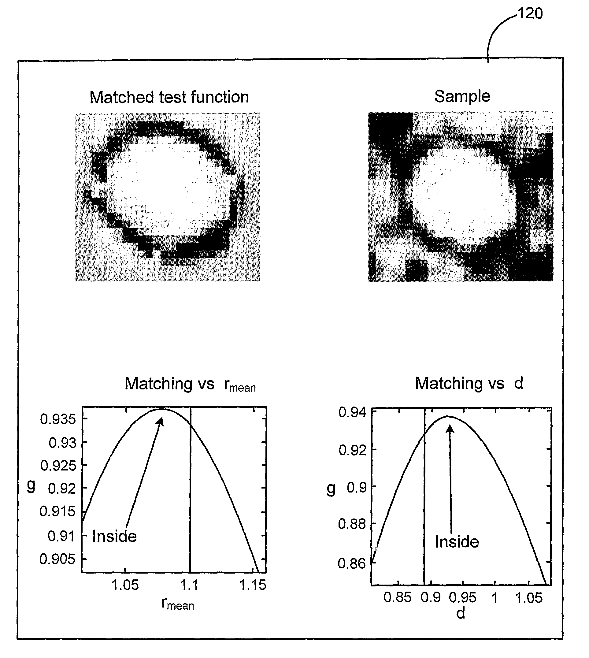
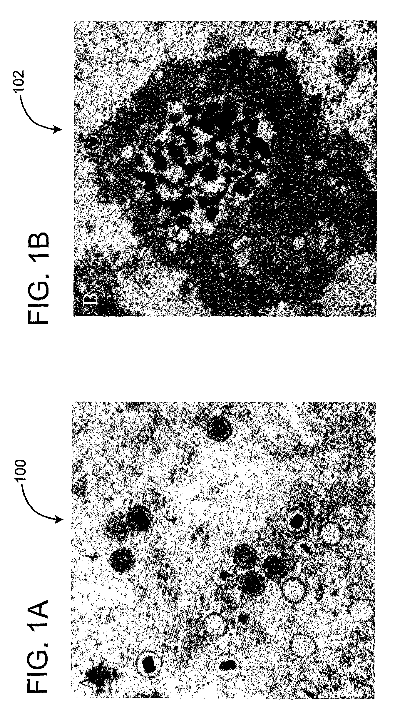
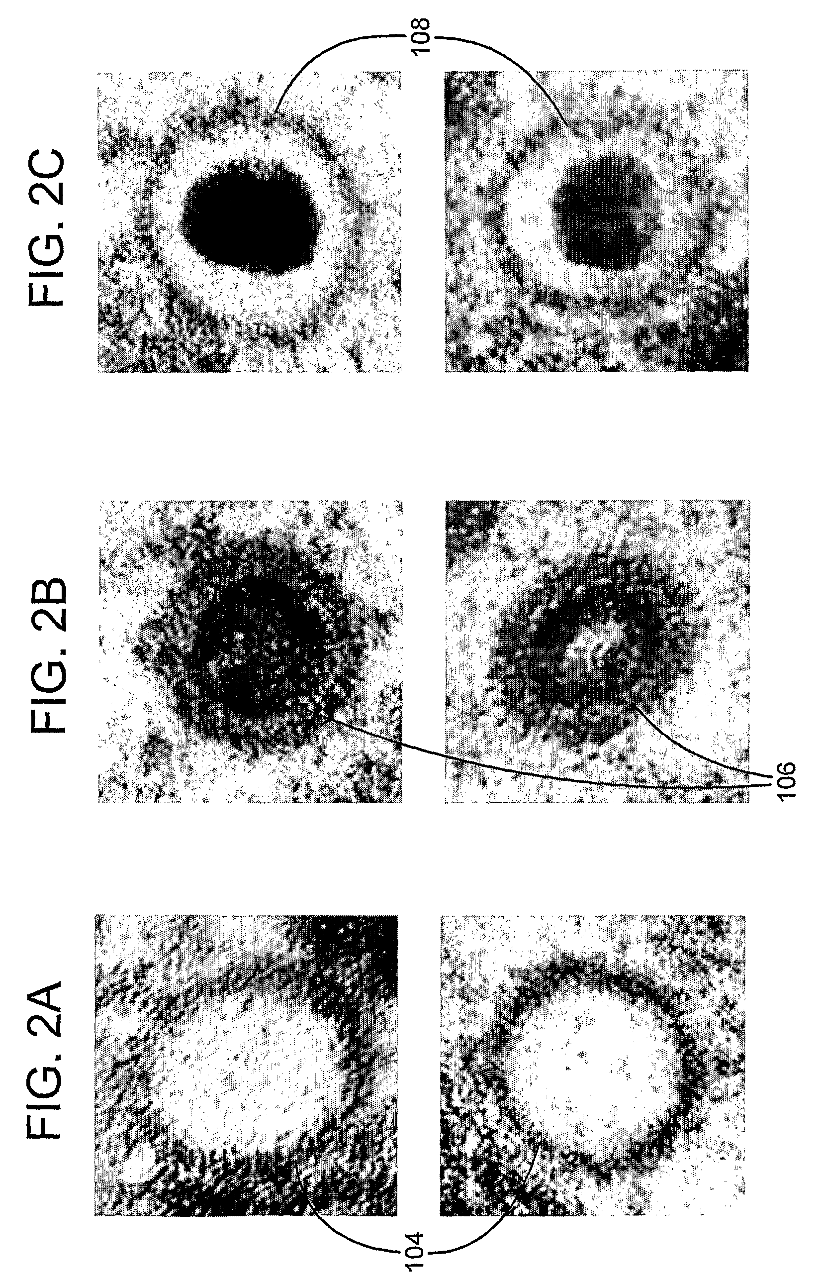
[0020]The development of an automated system to assist in the identification of virus particles in electron micrographs is herein described. As a model, fibroblasts have been used that are infected with human cytomegalovirus (HCMV) a virus of the β-herpes class. It should be understood that the herpes virus is only used as an illustrative example and the invention is not limited to the herpes virus. During infection with human cytomegalovirus, many different intermediate forms of the virus particle are produced. During assembly of the herpes virus, the host cell is forced to make copies of the viral genetic material and to produce capsids, a shell of viral proteins, which encase and protect the genetic material. Capsids are spherical structures that can vary with respect to size and symmetry and may, when mature be enveloped by a bi-layer membrane. The maturation of virus capsids is an important stage in virus particle production, and one that is frequently studied. However, their a...


PUM


Abstract

The method is for the identification and characterization of structures in electron micrographs. Structures in a first image are selected. The structures have a first shape type deformed in a first direction. The selected structures are transformed to a second shape type different from the first shape type. The transformed structures of the second shape type are used to form a plurality of templates. A new structure in a second image is identified. The new structure has the first shape type. The second shape type structure of each template is deformed in the first direction. It is determined which template is a preferred template that best matches the new structure.

Description

PRIOR APPLICATION[0001]This application is a U.S. national phase application based on International Application No. PCT / US2006 / 035758, filed 12 Sep. 2006, claiming priority from U.S. Provisional Patent Application No. 60 / 725,806, filed 12 Oct. 2005.FIELD OF THE INVENTION[0002]The present invention relates to identifying structures in images. In particular, the present invention provides a method and an arrangement of identification and classification of virus particles in textured electron micrographs.BACKGROUND OF THE INVENTION[0003]Virus assembly is an intricate process and a subject of intensive research. Viruses utilize a host cell to produce their progeny virus particles by undergoing a complex process of maturation and intracellular transport. This process can be monitored at high magnification utilizing electron microscopy, which allows visual identification of different types of virus particles in different cellular compartments. Important issues that remain to be resolved i...

Claims


Application Information

Patent Timeline
Patent Type & Authority Patents(United States)
IPC IPC(8): G06K9/62G06V10/772
CPCG06K9/6255G06K9/0014G06V20/695G06F18/28G06V10/772G06V10/32G06V10/40
Inventor HOMMAN, MOHAMMEDRYNER, MARTIN
Owner INTELLIGENT VIRUS IMAGING
Learn More