
Method and system for nodule feature extraction using background contextual information in chest x-ray images

a contextual information and nodule technology, applied in the field of chest x-ray image nodule feature extraction, can solve the problems of ineffective automatic nodule detection technique, difficult cognitively, and difficult deciphering of chest x-ray radiograph diagnosis of lung cancer, so as to improve the discriminating capability of extracted features and increase the effectiveness of distinguishing genuine nodules.

Active Publication Date: 2012-07-17
SIEMENS HEALTHCARE GMBH
View PDF14 Cites 3 Cited by
  • Summary
  • Abstract
  • Description
  • Claims
  • Application Information

AI Technical Summary

Benefits of technology

This approach enhances the discrimination capability of feature extraction, effectively distinguishing genuine nodules from false positives, even in complex backgrounds, improving the accuracy of nodule detection and reducing false positive errors.

Problems solved by technology

However, lung cancer diagnosis using chest x-ray radiographs can be very difficult cognitively.
Such diagnosis typically requires a radiologist to make decisions based on clues, which can be extremely difficult to decipher.
Unfortunately, an automatic nodule detection technique that is able to effectively cope with variations of chest x-ray images including different image characteristics, different types of nodules, and different background structures is not yet available.
Features that can effectively differentiate genuine nodules from similar background structures are difficult to extract.
Only a limited number of effective feature extraction techniques have been proposed.
However, a major disadvantage of such techniques is that they fail to incorporate sufficient nodule shape information when accumulating convergence evidence.
Matching filtering based techniques are able to remove a significant number of false positives, but have a limited capability in tolerating complex background structures.
They also lack the capability to handle weak nodules.
An important issue in nodule feature extraction is the localization of an effective region of interest.
However, nodule boundary localization is as difficult as nodule detection itself, if not more difficult.
The snake model approach is ineffective in handling background structures and weak nodules.
Practically, this is generally not feasible.
It is not practical to assume that adequate features are readily available.
Accordingly, relying solely on a learned classifier to dig out false positives may not be a feasible approach, since the features used by the learned classifier may not be sufficient in discriminating genuine nodules from false positives.
In addition, there are rarely enough representative genuine nodule samples available to enable robust learning.
The lack of discriminating capability of features and insufficient number of representative samples invariantly limit learning based techniques in practical applications.
Even though a significant amount of research has been concentrated on the issue of deriving effective features to differentiate nodules from false positives, the nodule feature extraction problem is far from solved.

Method used


Image

  • Method and system for nodule feature extraction using background contextual information in chest x-ray images
  • Method and system for nodule feature extraction using background contextual information in chest x-ray images
  • Method and system for nodule feature extraction using background contextual information in chest x-ray images

Examples


Embodiment Construction

[0020]The present invention is directed to a method for extracting nodule features using contextual background information in chest x-ray images. Embodiments of the present invention are described herein to give a visual understanding of the feature extraction method. A digital image is often composed of digital representations of one or more objects (or shapes). The digital representation of an object is often described herein in terms of identifying and manipulating the objects. Such manipulations are virtual manipulations accomplished in the memory or other circuitry / hardware of a computer system. Accordingly, is to be understood that embodiments of the present invention may be performed within a computer system using data stored within the computer system.

[0021]The foundations that current nodule detection techniques are based on are (1) nodules in chest x-ray radiographs are round-shaped blobs with some limited intensity difference (sometimes almost non-difference) from the res...


PUM

PropertyMeasurementUnit
shape analysisaaaaaaaaaa
propagation distanceaaaaaaaaaa
relative sizeaaaaaaaaaa

Abstract

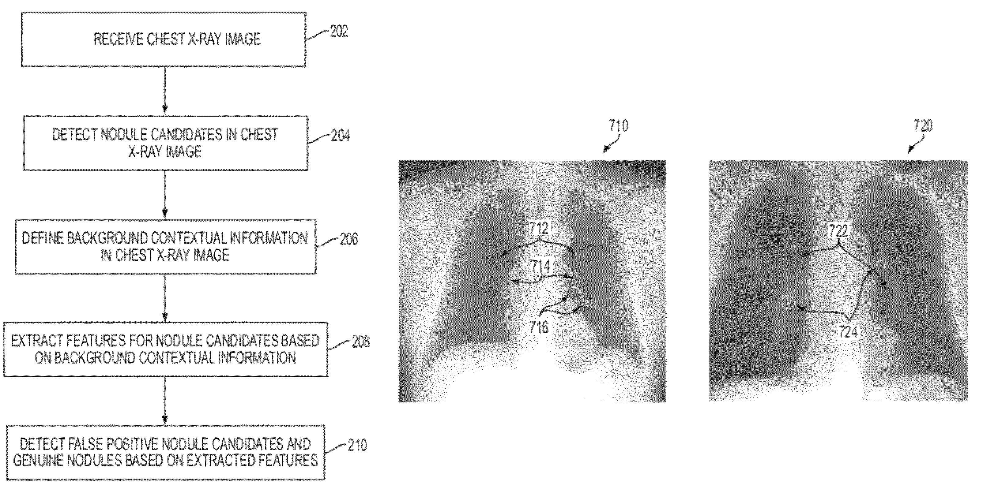
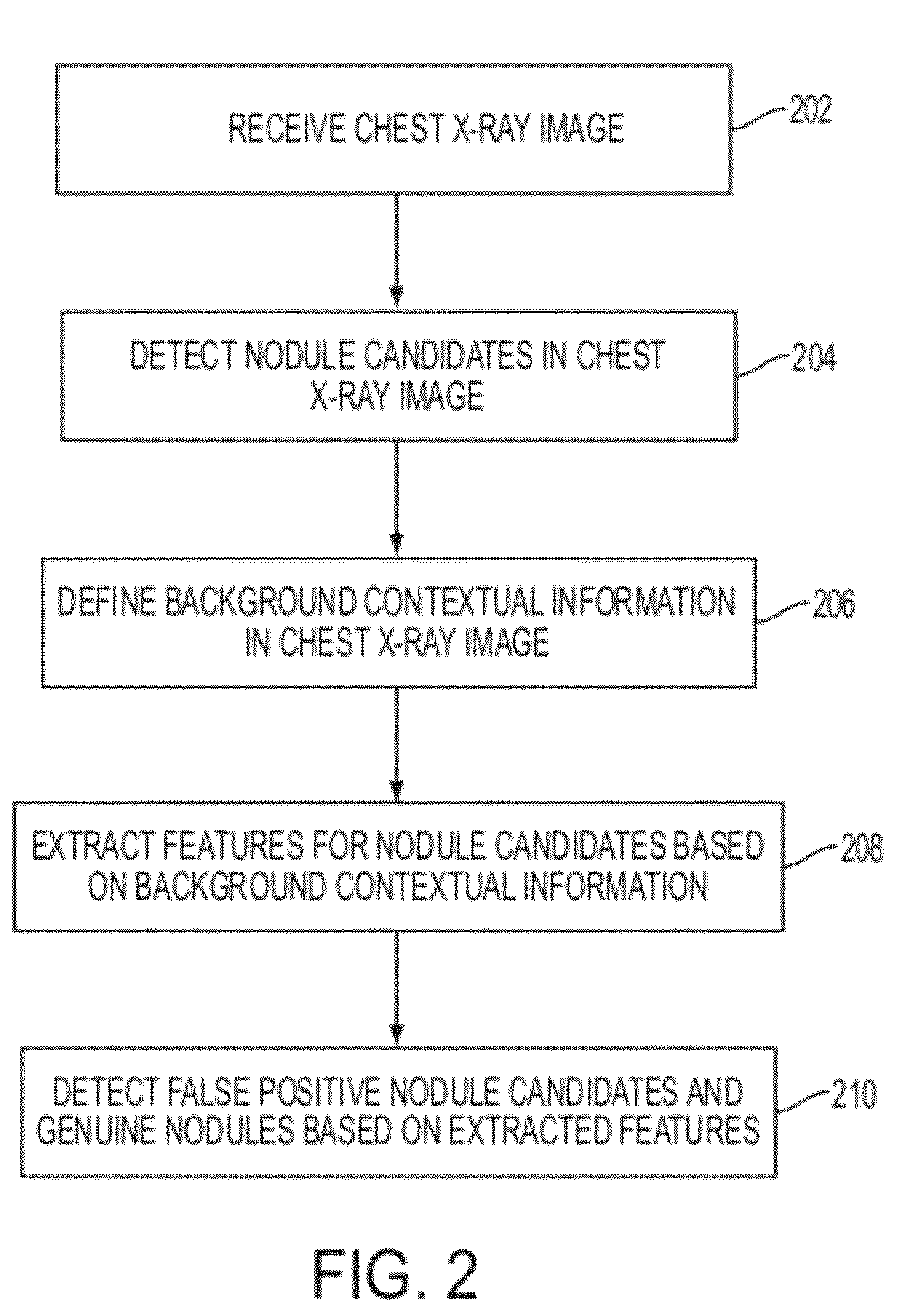
A method and system for nodule feature extract using background contextual information in chest x-ray images is disclosed. In order to detect false positives in nodule candidates for a chest x-ray image, background contextual information, such as contextual vessel tree information, is defined in the chest x-ray image. Features are extracted for each nodule candidate based on the background contextual information, and the extracted features are used to detect whether each nodule candidate is a false positive or a genuine nodule.

Description

[0001]This application claims the benefit of U.S. Provisional Application No. 60 / 980,866, filed Oct. 18, 2007, the disclosure of which is herein incorporated by reference.BACKGROUND OF THE INVENTION[0002]The present invention relates to detecting nodules in chest x-ray images, and more particularly, to nodule feature extraction in chest x-ray images.[0003]Lung cancer is a leading cause of all cancer deaths, and the survival rate can be significantly improved if it is detected in an early stage. Chest x-ray radiographs are a popular and cost effective way to perform initial examination and screening for lung cancer. In particular, chest x-ray radiographs are used to detect lung nodules. Nodules are small masses of tissue, which can form on various organs in the human body, such as the lungs. However, lung cancer diagnosis using chest x-ray radiographs can be very difficult cognitively. Such diagnosis typically requires a radiologist to make decisions based on clues, which can be extr...

Claims


Application Information

Patent Timeline
Patent Type & Authority Patents(United States)
IPC IPC(8): G06K9/46G01N23/04G06K9/78G06T7/60
CPCG06T7/0012G06K2009/00932G06K2209/051Y10S378/901G06T2207/30064G06K2209/053G06T2207/10116G06V40/14G06V2201/032G06V2201/031
Inventor HONG, LINSHEN, HONG
Owner SIEMENS HEALTHCARE GMBH
Learn More