
Devices and methods for securing tissue

a technology of inverted tissue and devices, applied in the direction of surgical staples, contraceptive devices, manufacturing tools, etc., to achieve the effect of extending the time between serosa and serosa healing

Active Publication Date: 2011-11-22
TENNESSEE MEDICAL INNOVATIONS INC
View PDF4 Cites 20 Cited by
  • Summary
  • Abstract
  • Description
  • Claims
  • Application Information

AI Technical Summary

Benefits of technology

The method effectively secures the diverticulum by compressing it serosa to serosa, reducing the risk of peritonitis and allowing for controlled healing by adjusting the compression ridge's sharpness for optimal healing time.

Problems solved by technology

A burst or leaky diverticulum results in the release of contaminants into the peritoneal cavity, potentially causing peritonitis.

Method used


Image

  • Devices and methods for securing tissue
  • Devices and methods for securing tissue
  • Devices and methods for securing tissue

Examples


Embodiment Construction

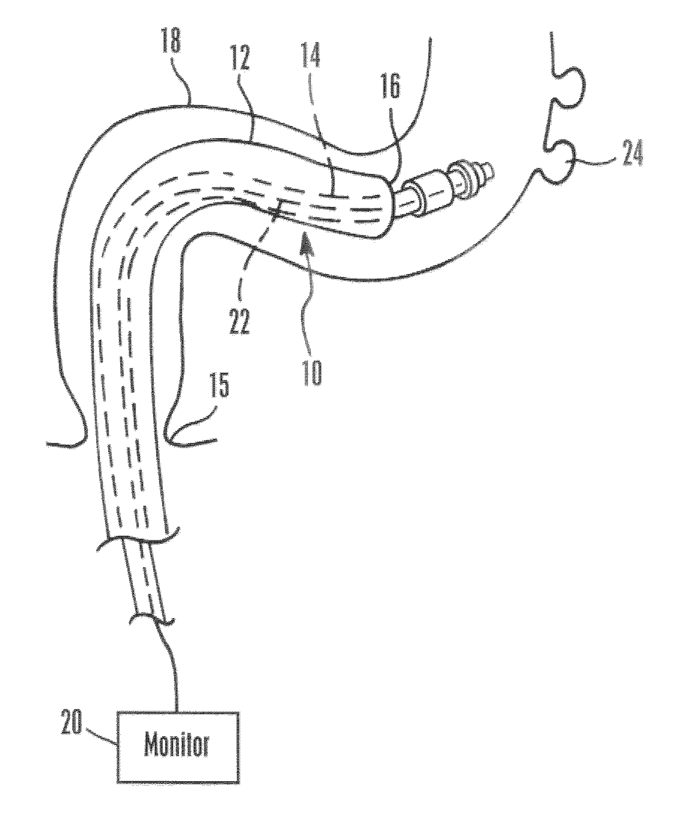
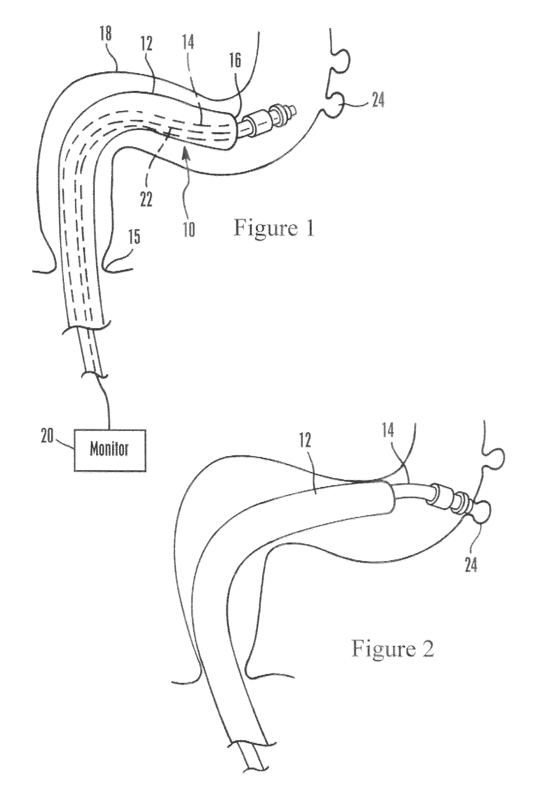
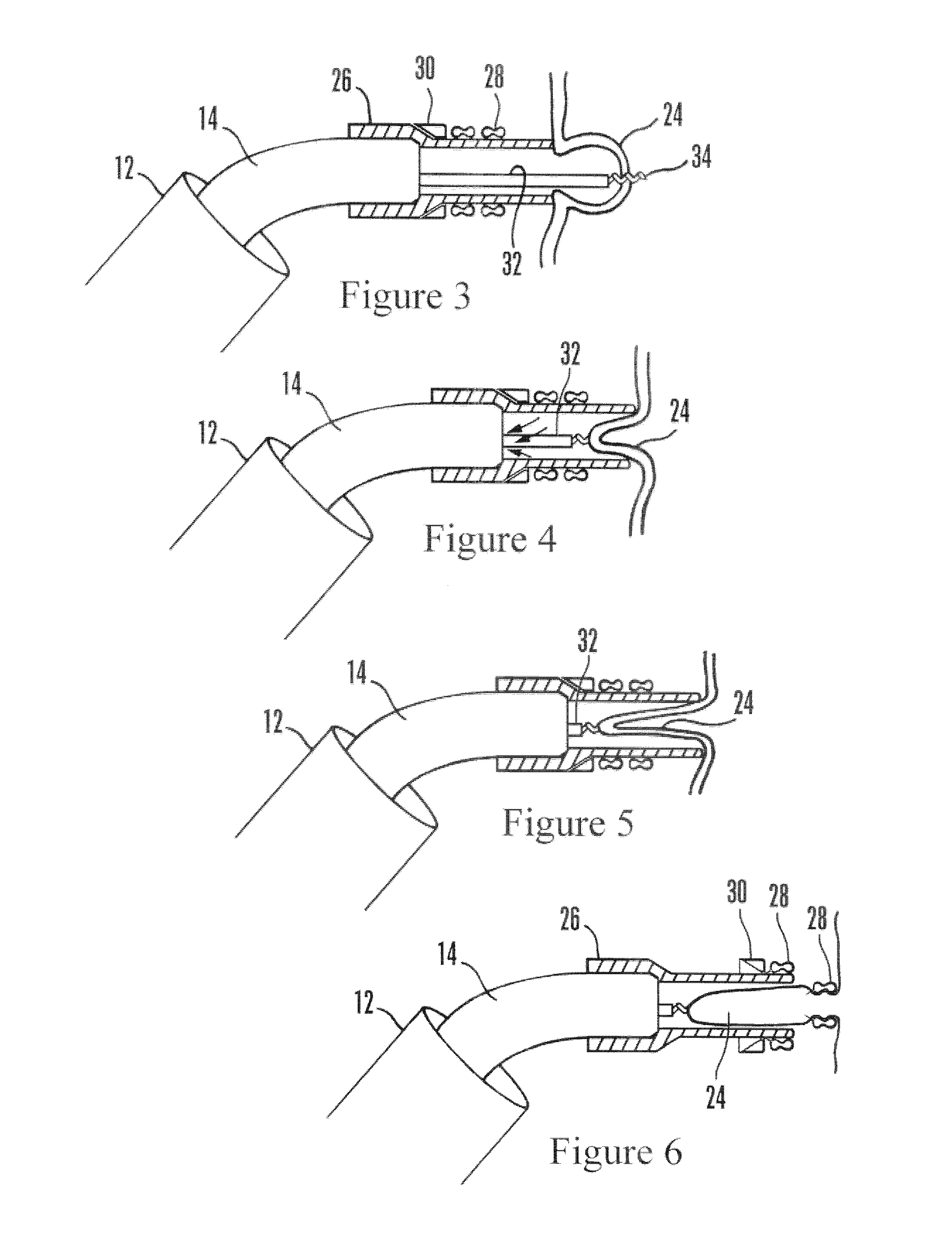
[0055]Referring initially to FIG. 1, a catheter assembly is shown, generally designated 10, that includes a flexible hollow overtube 12 fixedly or slidably holding one or more components such as but not limited to an endoscope such as a colonoscope 14 that may have plural working channels. The overtube 12 may be transparent plastic. The overtube 12 with colonoscope 14 is configured for being advanced into the patient through a natural orifice, such as the anus 15. The colonoscope 14 may extend from the open distal end 16 of the overtube 12 as shown to a colonoscope control handle (not shown) that is external to the patient. In this way, for example, images of the colon 18 from the colonoscope 14 can be presented on a monitor 20 to a surgeon. Accordingly, it will be appreciated that the colonoscope 14 bears one or more light guides 22 such as optical fibers for imaging the interior of the patient. The colonoscope 14 may extend through a working lumen of the overtube 12. Additional co...


PUM

PropertyMeasurementUnit
diameteraaaaaaaaaa
areaaaaaaaaaaa
axial dimensionaaaaaaaaaa

Abstract

A compression ring to grip and compress body structure such as diverticulum, hemorrhoids, and tissue adjacent a hole. A resilient ring-shaped body defines a compression channel, and one or more axially rigid elongated spikes extend from the body into the channel. The body defines a first axial segment surrounding the compression channel and a second axial segment surrounding the compression channel, with the spike being engaged only with the second axial segment. The first axial segment more tightly compresses the body structure than the second axial segment.

Description

[0001]This application claims priority from U.S. provisional patent application Ser. No. 60 / 982,083, filed Oct. 23, 2007 and 61 / 012,124, filed Dec. 7, 2007, both of which are incorporated herein by reference.FIELD OF THE INVENTION[0002]The present application relates generally to devices and methods for securing inverted tissue, and more particularly, though not exclusively, to securing inverted diverticulum serosa to serosa.BACKGROUND OF THE INVENTION[0003]Diverticulosis is an unfortunately common condition in which an area of the intestine bulges out into the peritoneal cavity to form a sac referred to as a “diverticulum”. Diverticulum are also formed when a point of weakness in the intestinal wall, e.g., where blood vessels take their entry, causes the muscular structure to divide and allows part of the inner mucosal membrane to be pushed out of the hole into the peritoneal cavity and become filled with fecal matter. Diverticulum may also be present in other parts of the gastroin...

Claims


Application Information

Patent Timeline
Patent Type & Authority Patents(United States)
IPC IPC(8): A61B17/12A61B17/122
CPCA61B17/12013A61B2017/00349A61B2017/00893Y10T29/53657A61B2017/12018A61B2017/306A61B2017/00296A61B2017/0641
Inventor NODA, WAYNE A.BELL, STEPHEN GRAHAM
Owner TENNESSEE MEDICAL INNOVATIONS INC
Learn More