Apparatus for obtaining ultrasonic image and method of obtaining ultrasonic image

an apparatus and ultrasonic image technology, applied in the field of can solve the problems of increasing the amount of data generated per unit of time, increasing the cost of the apparatus for obtaining ultrasonic images, and enlargement of hardware, so as to achieve the effect of suppressing an artifa

Inactive Publication Date: 2011-02-01
TOSHIBA MEDICAL SYST CORP
View PDF3 Cites 6 Cited by
  • Summary
  • Abstract
  • Description
  • Claims
  • Application Information

AI Technical Summary

Benefits of technology

[0026]An object of the invention is to provide an apparatus for obtaining an ultrasonic image, which scans a plurality of regions in accordance with a trigger signal based on an electrocardiographic waveform, capable of reducing a difference in time phases when scan data are obtained, and a method of obtaining an ultrasonic image.
[0028]In the vicinities of the boundary between the regions adjacent to each other, the scan data in which the time phases of the electrocardiographic waveform substantially coincide are obtained. Therefore, a difference in the time phases where the scan data are obtained can be reduced in the vicinities of the boundary. Accordingly, it is possible to suppress an artifact from being generated, the artifact being generated in a position corresponding to the boundary between the respective regions in the ultrasonic image.

Problems solved by technology

However, when scanning is performed by the two-dimensional ultrasonic probe, an amount of data generated per unit of time rapidly increases, compared with when scanning is performed by the one-dimensional ultrasonic probe.
Such enlargement of hardware significantly increases a cost of the apparatus for obtaining an ultrasonic image, which becomes a large obstacle to spread of the apparatus.
However, since the apparatus for obtaining an ultrasonic image according to the related art scans an ultrasonic beam in the same sub-scanning direction in the respective sub-volumes, the following problem occurs.

Method used


Image

  • Apparatus for obtaining ultrasonic image and method of obtaining ultrasonic image
  • Apparatus for obtaining ultrasonic image and method of obtaining ultrasonic image
  • Apparatus for obtaining ultrasonic image and method of obtaining ultrasonic image

Examples


second embodiment

[0140]Next, a second modification will be described with reference to FIGS. 13A to 13D. FIGS. 13A and 13B are schematic views showing a region scanned by an apparatus for obtaining an ultrasonic image according to the second modification. FIGS. 13C and 13D are schematic views illustrating the region scanned by the apparatus for obtaining an ultrasonic image according to the second modification and the scanning direction thereof, and are diagrams (top views) seen from the ultrasonic probe.

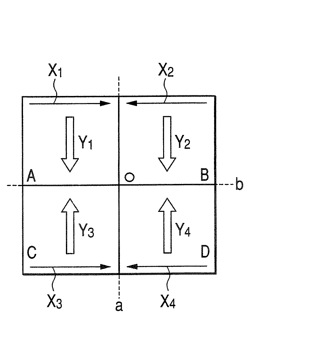
[0141]As shown in FIGS. 13A and 13B, the apparatus for obtaining an ultrasonic image according to the second modification scans an ultrasonic beam, with a circular conic region set to the scan region. Further, in the apparatus for obtaining an ultrasonic image according to the second embodiment, the regions equally-divided by dividing lines a and b are respectively set to the sub-volumes A to D, the dividing lines a and b passing through the center O of the circular conic scanning range and being or...


PUM


Abstract

A transmitting / receiving circuit drives an ultrasonic probe in accordance with a trigger signal based on an electrocardiographic waveform under the control of a control device so as to scan a plurality of regions, and obtains scan data for each region. During the scanning, the circuit causes the probe to scan the respective regions so that scan data in which time phases of the electrocardiographic waveform substantially coincide are obtained in the vicinities of the boundary between at least two adjacent regions among the regions. That is, in adjacent regions, the circuit causes the probe to scan the respective regions in the main or sub-scanning directions set to be reverse to each other. Further, an image processor generates ultrasonic image data of the range in which the regions are joined, on the basis of the scan data obtained by starting scanning at the same time phase.

Description

BACKGROUND OF THE INVENTION[0001]1. Field of the Invention[0002]The present invention relates to an apparatus for obtaining an ultrasonic image, which three-dimensionally transmits an ultrasonic wave into a subject and receives the reflected wave from the subject, thereby obtaining diagnosis information within the subject, and more particularly, to an apparatus for obtaining an ultrasonic image, which performs scanning by using an electrocardiographic signal (ECG signal).[0003]2. Description of the Related Art[0004]Recently, a so-called two-dimensional ultrasonic probe is being developed, in which ultrasonic transducers are two-dimensionally arranged so as to three-dimensionally transmit and receive an ultrasonic beam into and from a subject.[0005]As shown in FIG. 1, an apparatus for obtaining an ultrasonic image, provided with the two-dimensional ultrasonic probe, can transmit and receive an ultrasonic beam three-dimensionally. Therefore, the apparatus can scan the entire region of...

Claims


Application Information

Patent Timeline
Patent Type & Authority Patents(United States)
IPC IPC(8): A61B5/05
CPCA61B8/14A61B8/483G01S7/52085G01S7/52088G01S15/8993A61B8/543G01S15/8979
Inventor KOBAYASHI, TADAHARU
Owner TOSHIBA MEDICAL SYST CORP
Why Eureka
Try Eureka