
Systems and methods for viewing medical images

a medical image and system technology, applied in the field of management and viewing of medical images, can solve the problems of not allowing the viewer to accurately determine, cumbersome manual tracking of which portions of an image have been viewed at full resolution, and no system or method for automatically tracking the portions of an image that have been displayed at full resolution

Active Publication Date: 2010-02-09
MERATIVE US LP
View PDF71 Cites 72 Cited by
  • Summary
  • Abstract
  • Description
  • Claims
  • Application Information

AI Technical Summary

Problems solved by technology

As those of skill in the art will appreciate, manually tracking which portions of an image have been viewed at full resolution is cumbersome and may not allow the viewer to accurately determine when all relevant portions of the image have been viewed at full resolution.
Currently, there are no systems or methods for automatically tracking the portions of a medical image that have been displayed at full resolution, or for indicating those images for which all pixels have been presented on a display device at full resolution.

Method used


Image

  • Systems and methods for viewing medical images
  • Systems and methods for viewing medical images
  • Systems and methods for viewing medical images

Examples


Embodiment Construction

[0020]Embodiments of the invention will now be described with reference to the accompanying figures, wherein like numerals refer to like elements throughout. The terminology used in the description presented herein is not intended to be interpreted in any limited or restrictive manner, simply because it is being utilized in conjunction with a detailed description of certain specific embodiments of the invention. Furthermore, embodiments of the invention may include several novel features, no single one of which is solely responsible for its desirable attributes or which is essential to practicing the inventions herein described.

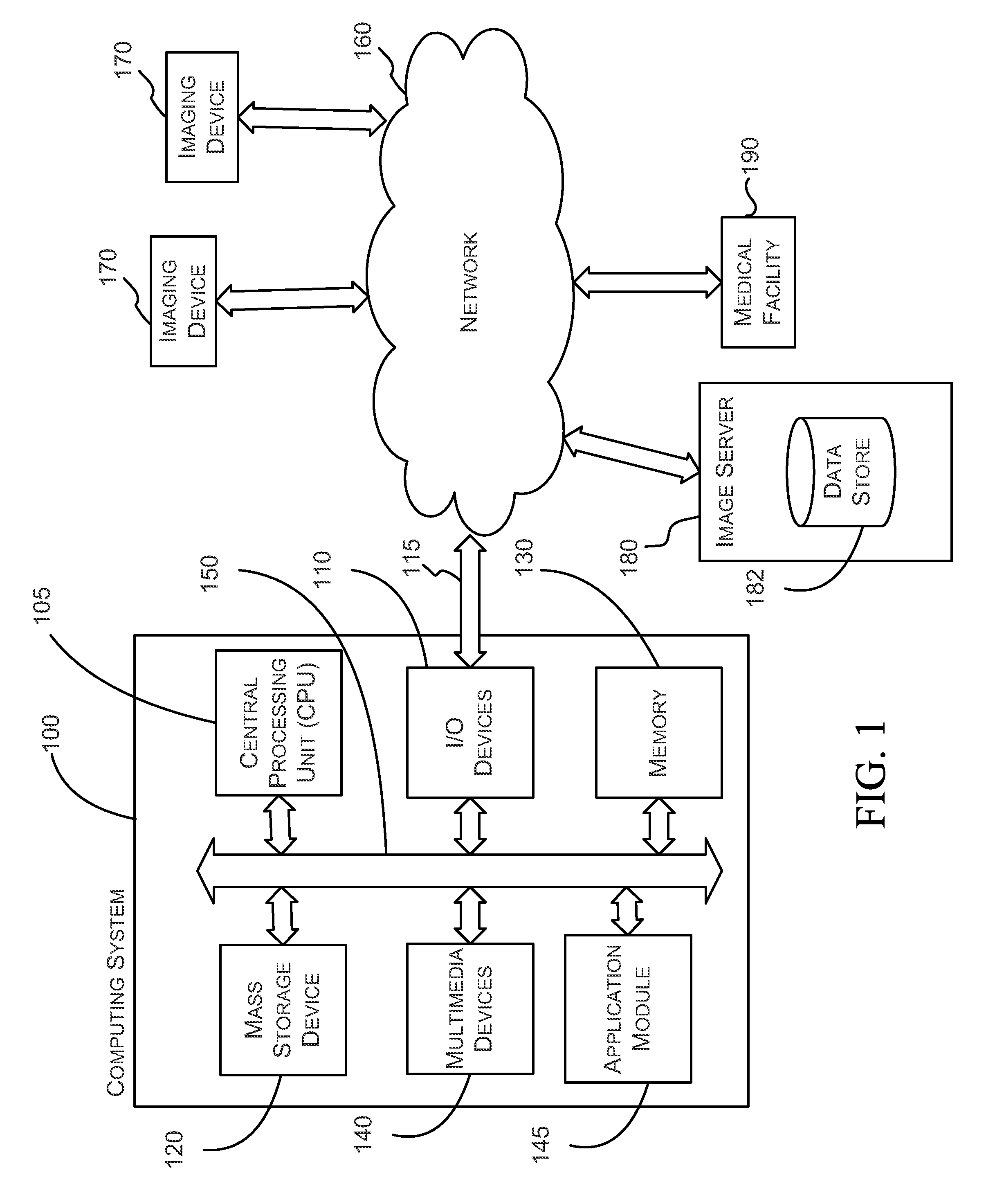
[0021]FIG. 1 is a block diagram of an exemplary computing system 100 in communication with a network 160 and various network devices. The computing system 100 may be used to implement certain systems and methods described herein. The functionality provided for in the components and modules of computing system 100 may be combined into fewer components and modu...


PUM


Abstract

For certain medical images, it is important and / or required that a user view all of a medical image at full resolution so that minute, but important, indicia in the medical image are not missed. A computing systems monitor the portions of the medical image that are displayed on the display device, notates those portions that have been displayed at full resolution (or other user-defined display parameters), and provides the user with information indicating portions that have not been viewed at full resolution and / or provides information indicating for which images of a multiple image examination full pixel display has been accomplished. The process reduces the possibility of missing an abnormality in a medical image due to the viewer not viewing a portion of the image at full resolution or using other user-defined display parameters.

Description

CROSS-REFERENCE TO RELATED APPLICATIONS[0001]This application claims priority under 35 U.S.C. § 119(e) to U.S. Provisional Application Ser. No. 60 / 625,690, filed on Nov. 4, 2004, which is hereby expressly incorporated by reference in its entirety.BACKGROUND OF THE INVENTION[0002]1. Field of the Invention[0003]This invention relates to management and viewing of medical images and, more particularly, to systems and methods of tracking which portions of medical images have been displayed using predetermined display parameters.[0004]2. Description of the Related Art[0005]Medical imaging is increasingly moving into the digital realm. This includes imaging techniques that were traditionally analog, such as mammography, x-ray imaging, angiography, endoscopy, and pathology, where information can now be acquired directly using digital sensors, or by digitizing information that was acquired in analog form. In addition, many imaging modalities are inherently digital, such as MRI, CT, nuclear m...

Claims


Application Information

Patent Timeline
Patent Type & Authority Patents(United States)
IPC IPC(8): G06K9/32G06K9/00G09G5/00
CPCG06F19/321G09G5/02G09G5/00G16H30/20G06F3/04845G06F3/167
Inventor REICHER, MURRAY A.FRAM, EVAN K.
Owner MERATIVE US LP
Learn More