
Methods and devices for the treatment of urinary incontinence

a technology for urinary incontinence and treatment methods, applied in the field of urinary incontinence treatment methods and devices, can solve the problems of urinary incontinence, risk of bone marrow infection and/or pubic osteitis, needles or carriers may wound large blood vessels in the operating region, etc., to facilitate transvaginal access of the pelvis, reduce the risk of accidental puncture, and blunt tissue dissection

Inactive Publication Date: 2009-05-05
BOSTON SCI SCIMED INC
View PDF249 Cites 93 Cited by
  • Summary
  • Abstract
  • Description
  • Claims
  • Application Information

AI Technical Summary

Benefits of technology

[0009]It is an object of this invention to provide instruments and methods for treating female stress urinary incontinence through transvaginal access and by blunt dissection. It is another object of this invention to provide instruments and methods for deploying sutures and / or slings in a surgical procedure, with reduced risk of accidental puncturing of the surgeon's fingers or injury to the patient's bladder and / or other abdominal organs.
[0010]In one another aspect, the present invention provides surgical instruments adapted for use in procedures to treat female urinary incontinence. The instrument includes a handle and a curved shaft that extends in a distal direction from the handle. The instrument is adapted to access interior tissue within a human body, such as the pelvic cavity. In one embodiment, the handle includes a friction-based gripping surface. A blunt tip is disposed at a distal end of the shaft for blunt dissection of tissue. The shaft is curved upward to facilitate transvaginal access of the pelvic cavity. In particular, the curved shaft and blunt tip allow the instrument to be moved through the endopelvic floor via blunt dissection, and to access retropubic tissue or suture attachment points such as Cooper's ligament or rectus fascia. A grasping mechanism, for grasping and cutting sutures or slings, may be located within a distal end portion of the shaft. The grasping mechanism is adapted to release a suture or a sling from the surgical instrument at a desired retropubic location. In an alternative embodiment, a hook deployment device may be attached to the surgical instrument through the grasping mechanism.

Problems solved by technology

Hypermobility is a condition in which the pelvic floor fails to properly support an area between the bladder neck and mid-urethra, thus permitting the urethra to descend from its normal anatomic position in response to increases in intra-abdominal pressure, resulting in urinary incontinence.
The introduction of such foreign objects into bone, however, presents a risk of bone marrow infection and / or pubic osteitis.
Because suspension needles and suture carriers include a sharp tip, there is a danger that the needle or carrier may wound large blood vessels present in the operating regions.
Sharp-tipped suspension needles and suture carriers also present a risk of puncturing or otherwise injuring the bladder and / or other abdominal organs.
Furthermore, the sharp end of the suspension needle or the suture carrier may cause puncture wounds to the surgeon's fingers, thus presenting a potential risk of infection to both the patient and the surgeon.
Such procedures involving “blind” dissection with sharp instruments is highly invasive and may cause significant trauma to the patient.

Method used


Image

  • Methods and devices for the treatment of urinary incontinence
  • Methods and devices for the treatment of urinary incontinence
  • Methods and devices for the treatment of urinary incontinence

Examples


Embodiment Construction

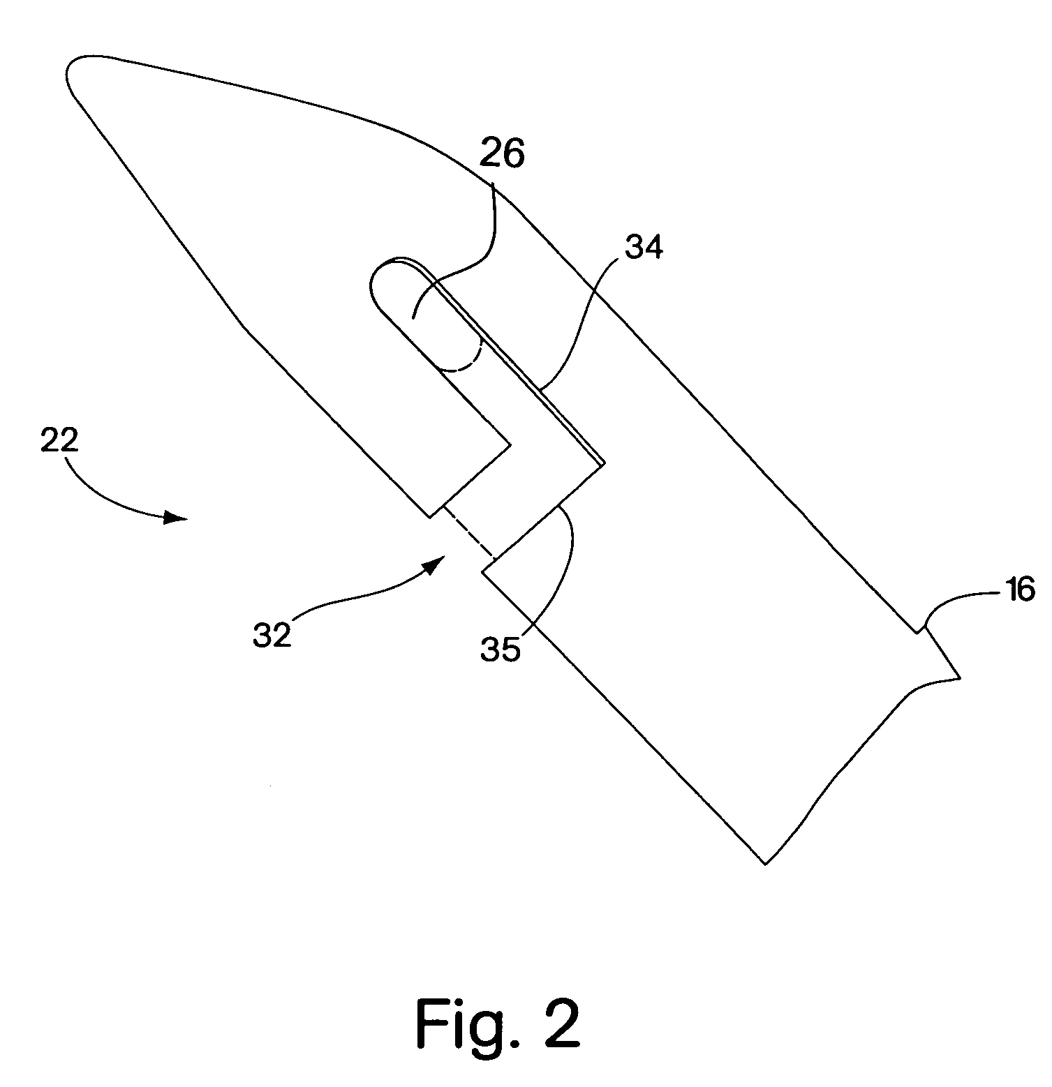
[0028]In overview, FIGS. 1 and 2 illustrate a surgical instrument 10 constructed according to the present invention for delivery of sutures or slings for the surgical treatment of female urinary incontinence. The curved surgical instrument 10 is constructed to transvaginally deliver sutures and / or slings to appropriate locations within the body to treat incontinence, without a need for dissection with sharp instruments.

[0029]The surgical instrument 10 is adaptable to be used in conjunction with a variety of tips and a variety of suture and / or sling deployment devices, providing the surgeon the flexibility to choose between different fixation methods.

[0030]A hook-type suture deployment device 50 is illustrated in FIGS. 3 and 4. The hook-type suture deployment device 50 provides for suture or sling attachment onto an anatomical support structure.

[0031]Referring to FIG. 1, a curved surgical instrument 10 according to the present invention comprises a handle 14 and a shaft 16 extending ...


PUM


Abstract

Methods and devices for treating female stress urinary incontinence are disclosed. The methods include transvaginally accessing the pelvic cavity and introducing a suburethral sling into the retropubic space. In some embodiments the ends of the sling are attached to an anatomical support structure. In other embodiments, the ends of the suburethral sling are not attached to an anatomical support structure. The devices include a surgical instrument for blunt dissection of the pelvic cavity which includes a curved shaft and a blunt distal end. A hook deployment device may optionally be attached to the surgical instrument.

Description

CROSS-REFERENCE TO RELATED APPLICATIONS[0001]This application claims the priority to and the benefit of U.S. provisional patent application Ser. No. 60 / 209,234 filed on Jun. 5, 2000, which is incorporated herein by reference in its entirety.TECHNICAL FIELD[0002]This invention relates to methods and devices for the treatment of female urinary incontinence.BACKGROUND INFORMATION[0003]Stress urinary incontinence in women may be caused by urethral hypermobility. Hypermobility is a condition in which the pelvic floor fails to properly support an area between the bladder neck and mid-urethra, thus permitting the urethra to descend from its normal anatomic position in response to increases in intra-abdominal pressure, resulting in urinary incontinence.[0004]Surgical procedures for stabilizing the urethra include suburethral slings and needle suspensions. In some sling procedures, a suburethral sling is placed around the posterior side of the urethra and the ends are attached to an anatomic...

Claims


Application Information

Patent Timeline
Patent Type & Authority Patents(United States)
IPC IPC(8): A61B17/04A61B17/00A61B17/06A61B17/28
CPCA61B17/0469A61B17/0483A61B17/06109A61B2017/00805A61B2017/06085A61B2017/2904
Inventor RIOUX, ROBERT F.
Owner BOSTON SCI SCIMED INC
Learn More