
Endoscopic subxiphoid surgical procedures

a subxiphoid and surgical technology, applied in the field of endoscopic surgical procedures, can solve the problems of inconvenient hospitalization, cumbersome space occupied by two sets of endoscopic equipment, etc., and achieve the effect of correcting regurgitation and reducing the size of the mitral annulus

Inactive Publication Date: 2007-09-04
ORIGIN MEDSYST +1
View PDF148 Cites 134 Cited by
  • Summary
  • Abstract
  • Description
  • Claims
  • Application Information

AI Technical Summary

Benefits of technology

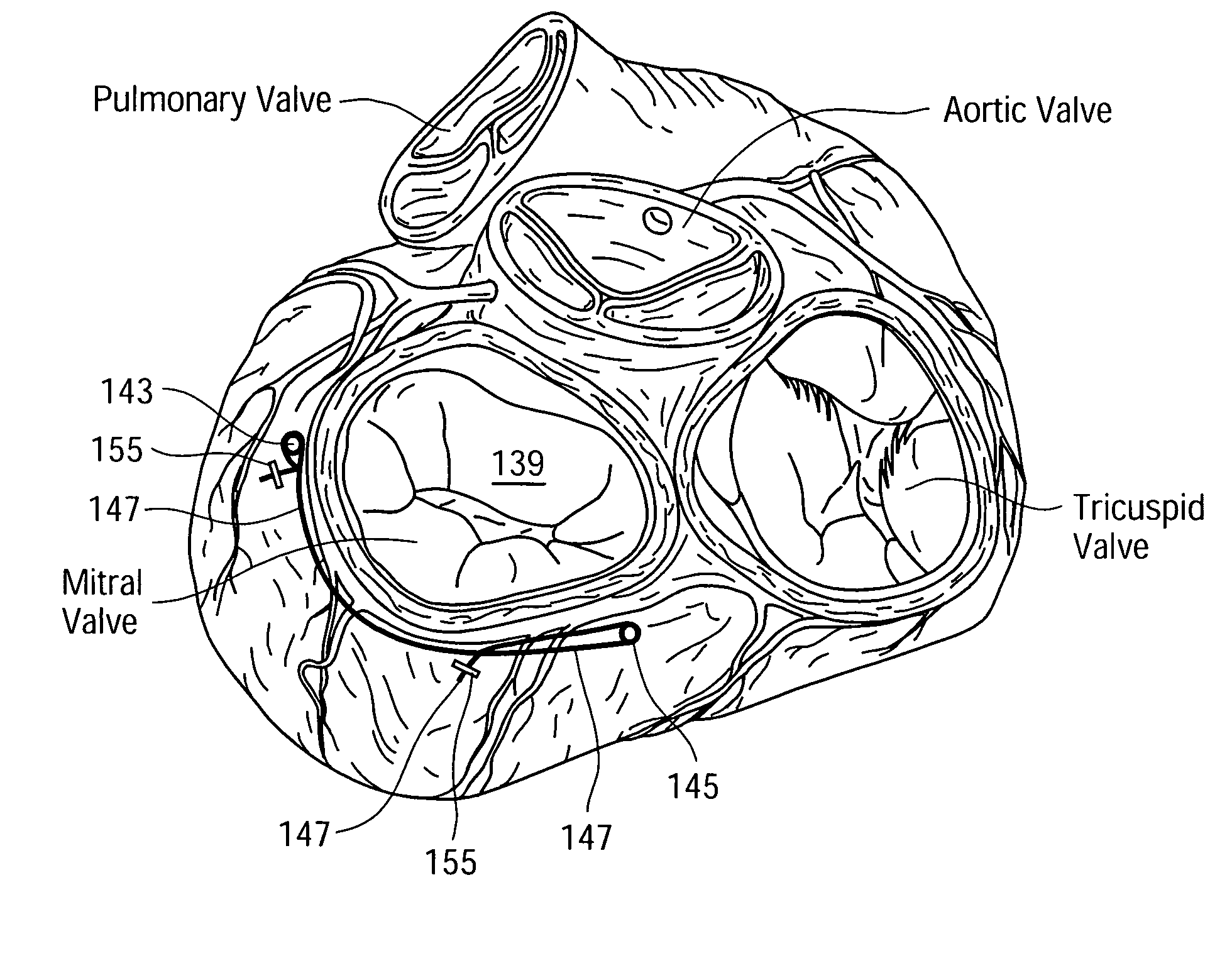
[0010]In accordance with another embodiment of the present invention, the endoscopic cannula is used to enter the pericardium from the subxiphoid approach to attach epicardial tacks and to tension the epicardium between tacks around the annulus of the mitral valve. Specifically, two or more tacks are placed on the epicardial surface near the mitral annulus. The tacks are connected by a suture or wire that may be tensioned to alter the shape and size of the annulus. The tacks may be placed immediately inferior to the left circumflex artery, in the area corresponding to the anterior aspect of the mitral annulus, and immediately inferior to the coronary sinus, in the area corresponding to the posterior aspect of the mitral annulus. The tacks may be helical or spiral titanium tacks of a type, for example, similar to tacks used to fixate prosthetic mesh in laparoscopic hernia repair. Two or more tacks may be inserted into the myocardium, and a suture or wire strand may be threaded through the portion of the tacks that is not embedded into the myocardium. The suture or wire contains loops spaced at varying distances for looping onto the tacks to adjust the amount of tension between the tacks. Tensioning the epicardium in this manner decreases the size of the mitral annulus and corrects the regurgitation due to annular dilation.

Problems solved by technology

The physical space occupied by two sets of endoscopic equipment is cumbersome in the operating room, and the expense is prohibitive to hospitals.

Method used


Image

  • Endoscopic subxiphoid surgical procedures
  • Endoscopic subxiphoid surgical procedures
  • Endoscopic subxiphoid surgical procedures

Examples


Embodiment Construction

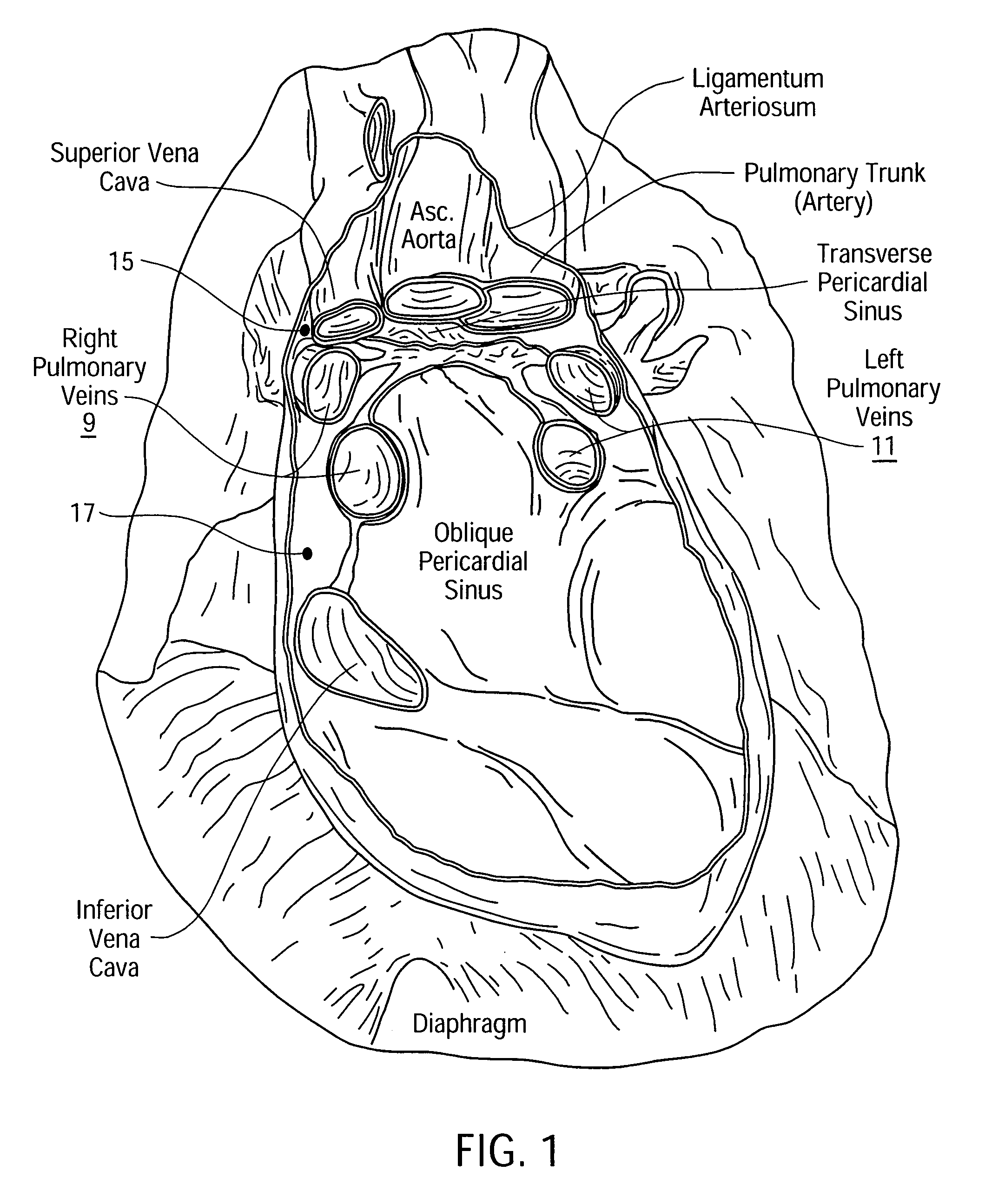
[0034]Referring now to FIG. 1, there is shown an anterior view of the interior of the pericardial sac (with the heart removed) that indicates the spatial orientations of various vessels including the right and left pulmonary veins 9, 11. A treatment for chronic atrial fibillation includes ablating cardiac tissue encircling the pulmonary veins 9, 11. This treatment may be accomplished in accordance with the present invention using an endoscopic cannula or probe via subxiphoid and thoracotomy access. Specifically, an ablation probe, as later described herein, or a tubular sheath therefor may be initially threaded around the pulmonary veins along a path 13 as indicated in FIG. 3, and the ablation probe may be subsequently advanced into position along the path 13 through the tubular sheath. In one embodiment, an endoscopic cannula enters the pericardium from a subxiphoid incision along a dissected channel in order to visualize the superior vena cava and place an illuminating clip, as la...


PUM


Abstract

Endoscopic subxiphoid surgical procedures and instruments facilitate translumination of tissue through the pericardium, and promote encircling an intrapericardial region with one or more tissue-ablating probes for ablating cardiac tissue substantially encircling the left and right pulmonary veins as a treatment for chronic atrial fibrillation. Such endoscopic subxiphoid surgical procedures and instruments also facilitate placement of epicardial tacks about the annulus of the mitral valve for supporting a tensioned suture or band that decreases the size of the mitral annulus to repair a regurgitant valve. Suction-oriented instruments facilitate temporary attachment to an organ to establish precise positioning on the organ during a surgical procedure.

Description

RELATED APPLICATIONS[0001]This is a continuation-in-part application of pending application Ser. No. 10 / 347,212, entitled “Apparatus and Methods for Endoscopic Surgical Procedures”, filed on Jan. 17, 2003 by Albert K. Chin, which is a continuation-in-part application of pending application Ser. No. 10 / 174,454, entitled “Releasable Guide And Method For Endoscopic Cardiac Lead Placement”, filed on Jun. 17, 2002 by Albert K. Chin, which is a continuation-in-part of pending application Ser. No. 10 / 140,309 entitled “Methods And Apparatus For Endoscopic Cardiac Surgery”, filed on May 6, 2002 by Albert K. Chin, et al., which is a continuation of pending application Ser. No. 09 / 635,721 entitled “Apparatus For Endoscopic Access”, filed on Aug. 9, 2000, which claims the benefit of provisional applications Ser. No. 60 / 148,130 filed on Aug. 10, 1999 and Ser. No. 60 / 150,737 filed on Aug. 25, 1999. This application is also a continuation-in-part application Ser. No. 09 / 779,715 entitled “Apparatus...

Claims


Application Information

Patent Timeline
IPC IPC(8): A61F2/00A61F2/24A61B1/01A61B17/00A61B17/06A61B17/22A61B17/30A61B17/32A61B17/34A61B18/00A61B18/14A61B19/00A61N1/05
CPCA61B17/00008A61B17/3403A61B17/3417A61B17/3421A61B19/201A61N1/0587A61B17/320068A61B17/3478A61B18/1482A61B19/54A61B2017/00243A61B2017/00247A61B2017/061A61B2017/22077A61B2017/306A61B2017/320044A61B2017/3445A61B2017/3488A61B2018/00291A61B2018/00392A61B2018/00982A61B2019/306A61B2019/462A61N2001/0578A61B90/11A61B2090/036A61B2090/062A61B90/39A61B2017/32007A61B2017/320069
Inventor CHIN, ALBERT K
Owner ORIGIN MEDSYST
Learn More