
Non-human animal exhibiting bone metastasis of tumor cells and method of screening for bone metastasis inhibitors

Inactive Publication Date: 2006-11-14
CHUGAI PHARMA CO LTD
View PDF2 Cites 3 Cited by
  • Summary
  • Abstract
  • Description
  • Claims
  • Application Information

AI Technical Summary

Benefits of technology

[0008]As a result of further studies based on the above findings, we have completed the present invention by finding that by using the above bone metastasis model, a novel agent for inhibiting and / or preventing bone metastasis can be screened.

Problems solved by technology

In both types, however, multi-organ metastasis is frequently observed at the time of diagnosis.
In particular, bone metastasis causes various severe symptoms by inducing pathological fracture, compression of spinal cord, and hypercalcemia, and reduces the quality of life of the patients (Iguchi, H. et al., Cancer Res. 56:4040–4043, 1996).
However, intracardiac injection of tumor cells does not seem to be technically easy, and is associated to some extent with sudden death of the animal.
Therefore, intracardiac injection of tumor cells imposes a substantial burden on animals, causing ethical and economic problems.
As a result, a simple method with good reproducibility of screening a novel agent to solve the important problem, that is, inhibition and / or prevention of bone metastasis, has not yet been developed.

Method used


Image

  • Non-human animal exhibiting bone metastasis of tumor cells and method of screening for bone metastasis inhibitors
  • Non-human animal exhibiting bone metastasis of tumor cells and method of screening for bone metastasis inhibitors
  • Non-human animal exhibiting bone metastasis of tumor cells and method of screening for bone metastasis inhibitors

Examples


example 1

[0107]Pattern of Metastasis Produced by Human Lung Cancer Cell Lines in NK-cell Depleted SCID Mice

[0108]We examined the pattern of multi-organ metastasis produced by 8 different human lung cancer cell lines in NK-cell depleted SCID mice. NK-cell depleted SCID mice were injected intravenously through tail vein into the mice with 1–5×10⁶ tumor cells, and were sacrificed on the day after the indicated periods, and the number of metastatic colonies into the lungs, livers, kidneys and lymph nodes were counted All recipient mice developed tumor lesion and many of the mice became morbid by the time of sacrifice (Table 1).

[0109]

TABLE 1Pattern of Metastasis Produced by HumanLung Cancer Cell Lines in NK-Cell Depleted SCID MiceDay ofNumber of Metastases; median (Range)Cell LineSacrificeBoneLungsLiverKidneysLymph NodesSquamous cell carcinomaRERF-LC-AIaday 42All 0All 067 (38–100)19 (15–30) 4 (0–18)AdenocarcinomaPC-14aday 28All 0>100 3 (1–7) 5 (3–13) 1 (0–3)A549aday 56All 0>100 1 (0–2)All 010 (4–...

example 2

[0111]X-ray and Histological Analysis of Bone Metastasis Produced by SBC-5 Cells

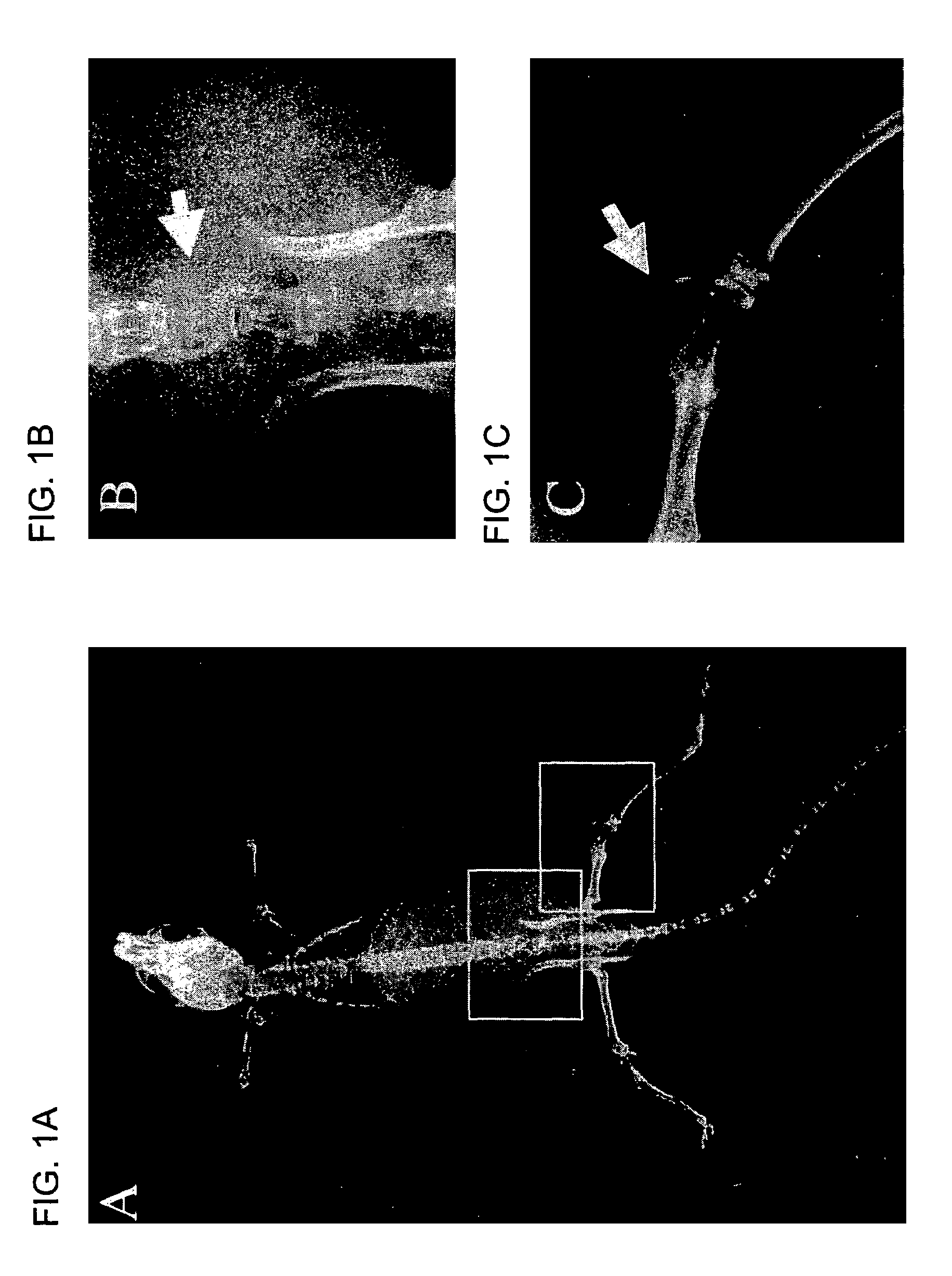
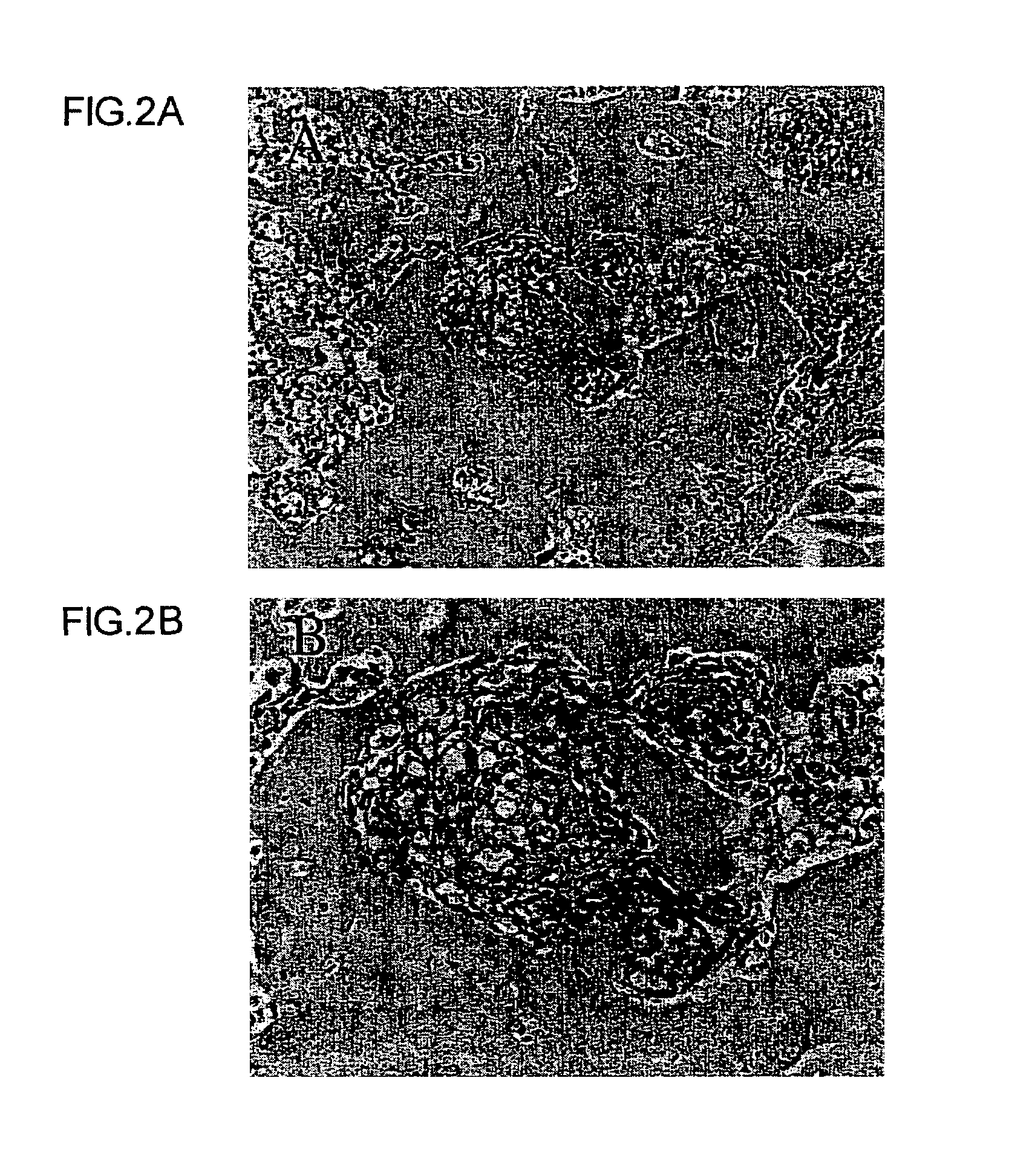
[0112]Bone metastases obtained in Example 1 were detected by X-ray photography. Multiple bone metastases were reproducibly developed in the mice injected intravenously with SBC-5 cells and bone metastasis lesions were detected as radiolucent lesions on X-ray photograph (FIG. 1A), indicating osteolytic bone metastases mainly in the spine (FIG. 1B) and bone of extremities (FIG. 1C). Histological analysis shows that these lesions consist of tumor cells with multi-nucleated cells (FIGS. 2A and B). The mice with these lesions had paralysis of hind legs and urinary retention with enlarged bladder, probably due to pathological fracture and / or compression of spinal cord caused by bone metastasis.

example 3

[0113]Effect of Tumor-cell Number on Bone Metastasis in NK-cell Depleted SCID Mice

[0114]To determine the optimal experimental conditions for bone metastasis, we injected various numbers of SBC-5 cells into NK-cell depleted SCID) mice. When the mice became moribund, the mice were sacrificed and the number of bone metastasis was determined by X-ray photography. The number of visceral organs was determined macroscopically. The number of bone metastases, as well as visceral metastases, depended on the number of cells injected (Table 2). Based on these results, 1×10⁶ SBC-5 cells were injected in subsequent experiments.

[0115]

TABLE 2Pattern of Multiple Organ Metastases Produced by SBC-5 Cells in NK-Cell Depleted SCID MiceNumber ofDay ofBoneLungsLiverKidneysLymph NodesInjectionSacrificeInc.aMed.bRangeInc.MedRangeInc.MedRangeInc.MedRangeInc.MedRange1 × 1051213 / 530–60 / 50All 05 / 530 7–450 / 50All 02 / 530–55 × 105495 / 561–82 / 530–105 / 52514–503 / 520–4 3 / 520–31 × 106355 / 542–55 / 553–185 / 54725–585 / 521–3 5 / ...


PUM


Abstract

The present invention provides a non-human bone metastasis model animal in which tumor cells capable of inducing bone metastasis by peripheral administration have been introduced, and a method for producing the animal. The bone metastasis model animal in the present invention may be useful for understanding the biology of bone metastasis and developing novel therapeutic strategies for lung cancer patient with multi-organ metastases, including bone metastasis. The present invention also provides a method of screening an agent for inhibition and / or prevention of bone metastasis of tumor cells using the non-human bone metastasis model animal.

Description

[0001]This application is a continuation-in-part of U.S. application Ser. No. 09 / 864,364, filed May 25, 2001 now abandoned, and Japanese Patent Application 2001-140538, filed May 10, 2001, which are herein incorporated by reference for any purpose.FIELD OF THE INVENTION[0002]The present invention relates to a non-human animal exhibiting bone metastasis of tumor cells, a method for producing the animal, and a method of screening an agent for inhibiting and / or preventing bone metastasis of tumor cells. In particular, the present invention relates to the animal in which tumor cells capable of inducing bone metastasis by peripheral administration have been introduced, and a method for producing the animal. The invention also relates to a method of screening a novel agent, comprising the steps of producing a non-human model animal exhibiting multi-organ metastasis of tumor cells, including bone metastasis, and treating the model animal with the novel agent.BACKGROUND OF THE INVENTION[000...

Claims


Application Information

Patent Timeline
IPC IPC(8): A01K67/00A01K67/027
CPCA01K67/0271
Inventor SONE, SABUROMIKI, TOYOKAZU
Owner CHUGAI PHARMA CO LTD
Learn More