
Method of securing tissue

a tissue and anastomosis technology, applied in the direction of surgical staples, applications, mechanical equipment, etc., can solve the problems of recurrence of angina, myocardial infarction and death, and high cost of such procedures, and achieve the effect of reducing the risk of anastomosis

Inactive Publication Date: 2006-04-11
BOSTON SCI SCIMED INC
View PDF101 Cites 178 Cited by
  • Summary
  • Abstract
  • Description
  • Claims
  • Application Information

AI Technical Summary

Benefits of technology

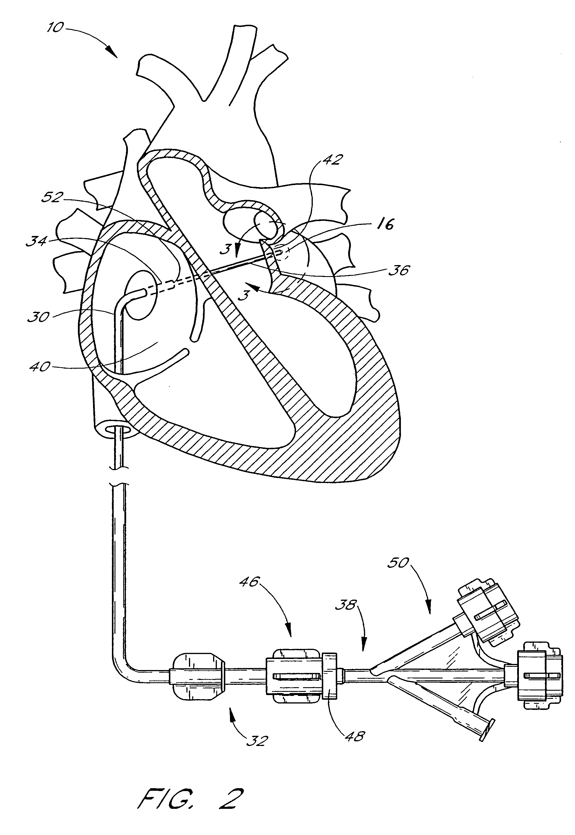
The present invention provides a method for attaching a tubular graft to a vessel wall using tissue anchors. The method involves positioning a graft within a vessel and a tissue anchor deployment catheter within the graft. The anchors are then advanced into the vessel wall to secure the graft. The invention also provides an anastomosis catheter for creating a connection between two tubular structures. The catheter has a tissue anchor support that can be moved from an axial to an inclined position to secure the graft to the vessel wall. The invention also includes a method for tacking a graft to a vessel wall using tissue anchors. Overall, the invention provides a reliable and effective method for attaching a graft to a vessel wall.

Problems solved by technology

While the typical coronary bypass procedure favorably affects the incidence and severity of angina in patients with coronary artery disease, a variety of risks are associated with such procedures.
Furthermore, the vein grafts deteriorate over time, thereby resulting in the recurrence of angina, myocardial infarction and death.
In addition, the costs of such procedures are relatively high and the patient recovery relatively long.
While vascular anastomosis can be effective, and sometimes life-saving procedures, traditionally available techniques have been associated with a number of complications.
Such operations are traumatic to the patient, involve a lengthy recovery, and a relatively high risk of infection or other complications.
As a result, these operations typically require large numbers of sutures or staples to close the incision and 5 to 10 wire hooks to keep the severed sternum together.
Furthermore, such procedures leave an unattractive scar and are painful to the patient.
Most patients are out of work for a long period after such an operation and have restricted movement for several weeks.
Such surgery often carries additional complications such as instability of the sternum, post-operative bleeding and mediastinal infection.
In this regard, limited open chest techniques have been developed in which the coronary bypass is carried out using an abdominal (subxyphoid) approach or, alternatively, a “Chamberlain” incision (an approximately 8 cm incision at the sternocostal junction), thereby lessening the operating area and the associated complication rate.
Nevertheless, each of these techniques is thoracotomic, requiring an incision to be made in the chest wall through which conventional surgical instruments are introduced to perform conventional coronary bypass surgery.
Suturing of vascular structures, however, is a tedious and time consuming process.
Furthermore, current suturing techniques are not possible using transluminal access, and are not readily adapted for endoscopic use, where the surgeon's freedom of access and movement are limited.
Due to the size of these devices, however, they are not easily adapted for use with vascular organs in general, and particularly not for transluminal or endoscopic techniques.
A transluminal approach is thus not readily possible using this technique.

Method used


Image

  • Method of securing tissue
  • Method of securing tissue
  • Method of securing tissue

Examples


Embodiment Construction

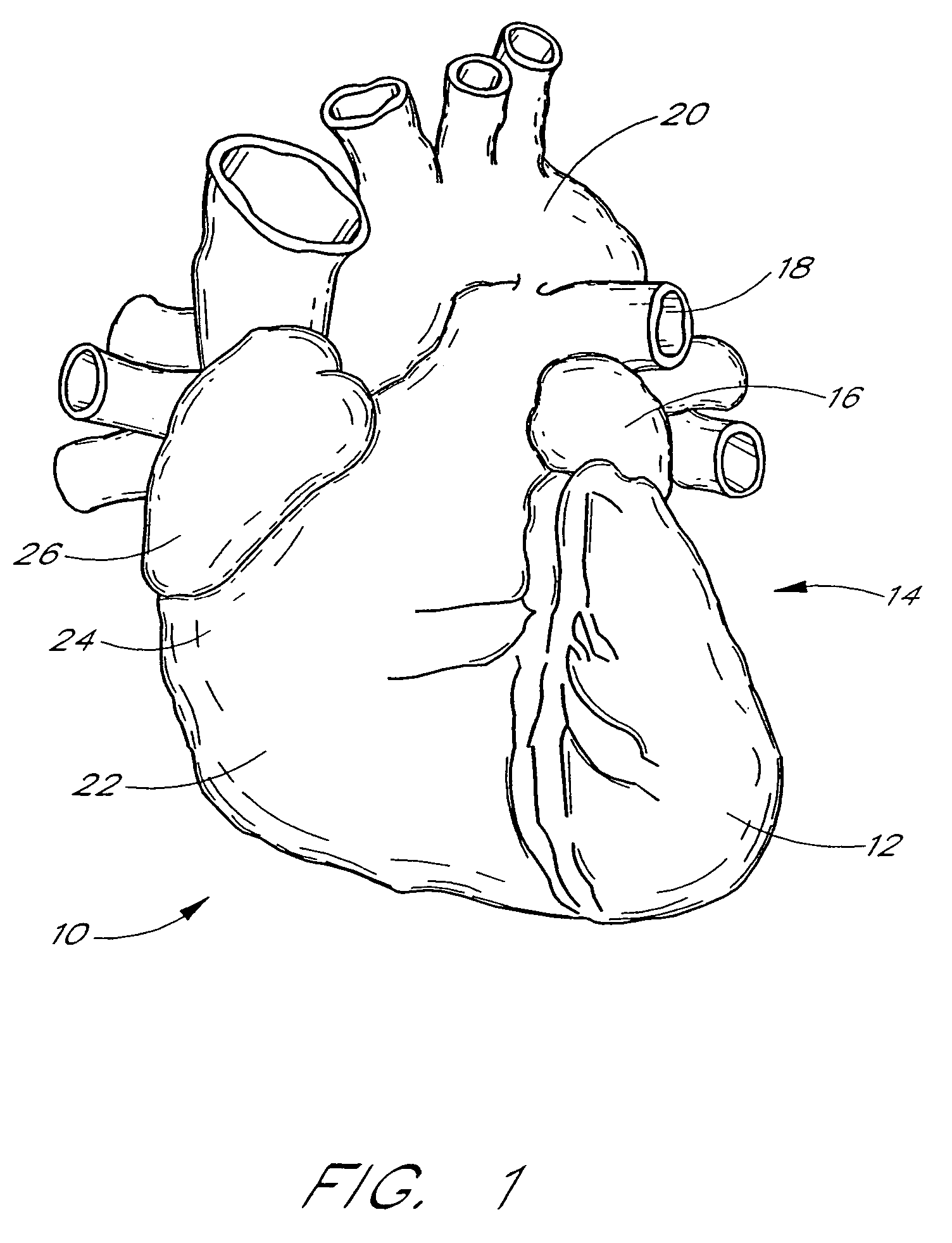
[0043]For simplicity, the present invention will be described primarily in the context of a left atrial appendage closure procedure, and as modified for use in tissue-to-tissue or synthetic graft-to-tissue anastomosis. As used herein the term “anastomosis” shall include securing a tubular synthetic graft within a vessel, such as to span an aneurysm, as well as the end to end and end to side orientation discussed in the Background of the Invention. However, the device and methods herein are readily applicable to a wider variety of closure or attachment procedures, and all such applications are contemplated by the present inventors. For example, additional heart muscle procedures such as atrial septal defect closure and patent ductus arteriosis closure are contemplated. Vascular procedures such as isolation or repair of aneurysms, anastomosis of vessel to vessel or vessel to prosthetic tubular graft (e.g., PTFE or Dacron tubes, with or without wire support structures as are well known...


PUM


Abstract

Disclosed is an anastomosis catheter, for achieving a tissue to tissue or synthetic graft to tissue attachment. The catheter includes a plurality of deployable tissue anchors, which may be laterally deployed into surrounding tissue. The anchors may be used to achieve end to end or end to side anastomoses. Methods are also disclosed.

Description

[0001]This application is a continuation of application Ser. No. 09 / 903,970 filed Jul. 11, 2001, now U.S. Pat. No. 6,746,472, which is a divisional of application Ser. No. 09 / 482,986 filed on Jan. 11, 2000, now U.S. Pat. No. 6,328,727, which is a continuation-in-part of application Ser. No. 09 / 399,521, filed Sep. 20, 1999, now U.S. Pat. No. 6,231,561, the disclosure of which is incorporated in its entirety herein by reference.BACKGROUND OF THE INVENTION[0002]The present invention relates to methods and devices for performing anastomosis. More particularly, the present invention relates to methods and devices for performing tissue-to-tissue or synthetic graft-to-tissue vascular anastomosis under either direct or transluminal access.[0003]Anastomosis is the union or joinder of one hollow vessel or structure to another so that the interior of the vessels communicate with one another. There are generally two types of vascular anastomosis: end-to-end and end-to-side. In an end-to-end ana...

Claims


Application Information

Patent Timeline
Patent Type & Authority Patents(United States)
IPC IPC(8): A61M31/00A61B17/00A61B17/04A61B17/064A61B17/11A61F2/24A61F2/82
CPCA61B17/00234A61B17/0401A61B17/12122A61B17/0057Y10S606/907A61B17/064A61B17/0644A61B2017/00243A61B2017/00575A61B2017/00579A61B2017/00632A61B2017/0409A61B2017/0412A61B2017/0414A61B2017/0417A61B2017/042A61B2017/0437A61B2017/0445A61B2017/0454A61B2017/0458A61B2017/0464A61B2017/0472A61B2017/0477A61B2017/048A61B2017/0488A61B2017/0647A61F2/24A61F2/2427A61F2/2487A61B2017/00292A61B2017/0645A61B2017/00668
Inventor FRAZIER, ANDREW G. C.LESH, MICHAEL D.ROUE, CHAD C.VAN DER BURG, ERIK J.
Owner BOSTON SCI SCIMED INC
Learn More