
Image diagnosis assistance apparatus, endoscope system, image diagnosis assistance method , and image diagnosis assistance program

an image diagnosis and assistance technology, applied in the field of image diagnosis assistance apparatus, an endoscope system, an image diagnosis assistance method, etc., can solve the problems of reducing the motivation of operators, high risk of oversight, and high need for reporting

Pending Publication Date: 2022-05-19
FUJIFILM CORP
View PDF0 Cites 0 Cited by
  • Summary
  • Abstract
  • Description
  • Claims
  • Application Information

AI Technical Summary

Benefits of technology

[0020]As described above, the image diagnosis assistance apparatus, the endoscope system, the image diagnosis assistance method, and the image di

Problems solved by technology

For several seconds immediately after a region of interest comes into sight, a viewpoint frequently changes and the risk of oversight is high, and thus necessity for reporting is high.
Once an operator has found a region of interest and started detailed observation, reporting may disturb the observation, reduce the operator's motivation to perform an operation

Method used


Image

  • Image diagnosis assistance apparatus, endoscope system, image diagnosis assistance method , and image diagnosis assistance program
  • Image diagnosis assistance apparatus, endoscope system, image diagnosis assistance method , and image diagnosis assistance program
  • Image diagnosis assistance apparatus, endoscope system, image diagnosis assistance method , and image diagnosis assistance program

Examples


first embodiment

Configuration of Endoscope System

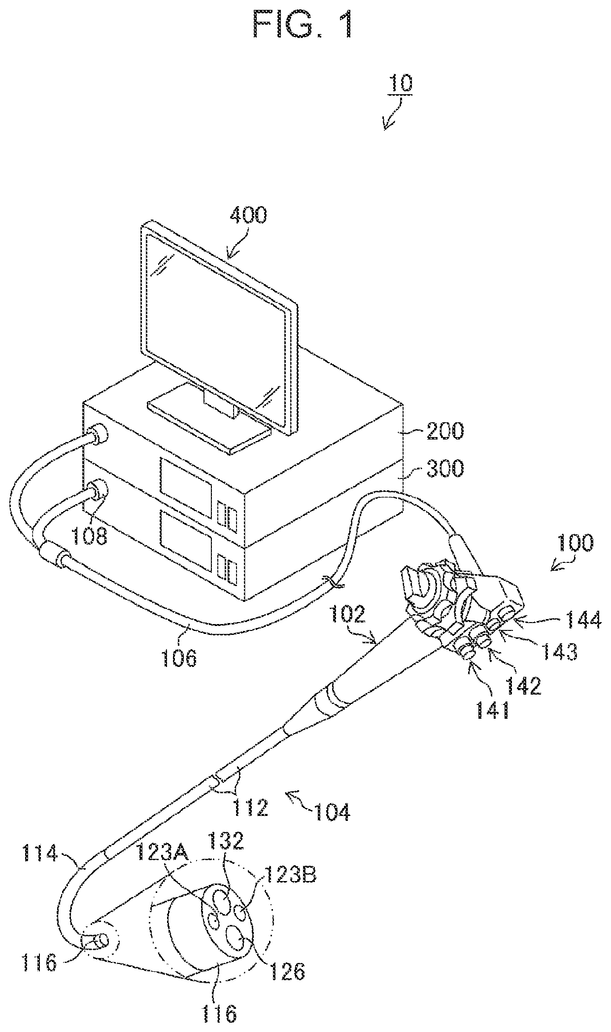
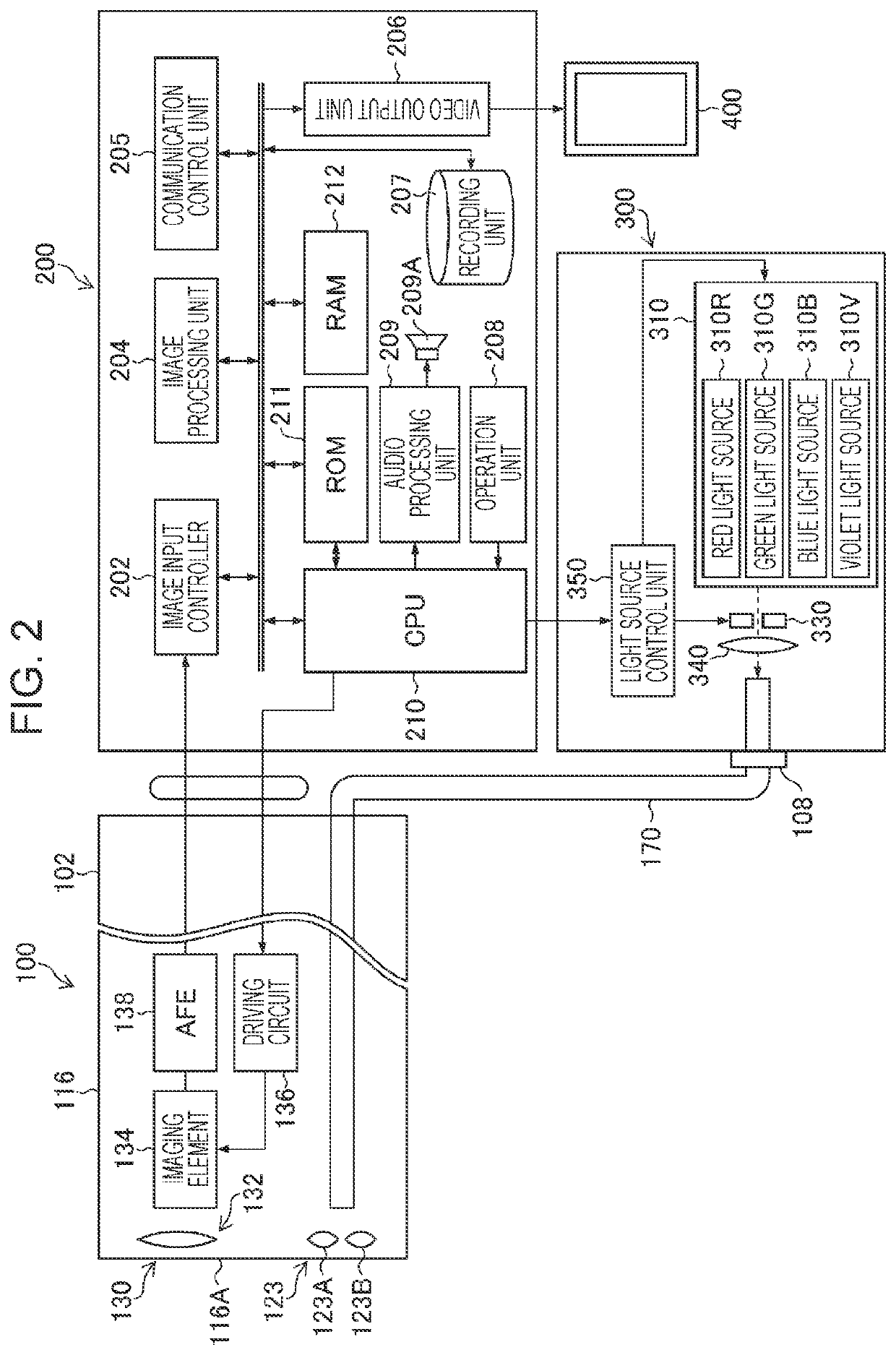
[0037]FIG. 1 is an external appearance diagram of an endoscope system 10 (an image diagnosis assistance apparatus, a medical image processing apparatus, an endoscope system), and FIG. 2 is a block diagram illustrating the configuration of a main part of the endoscope system 10. As illustrated in FIGS. 1 and 2, the endoscope system 10 is constituted by an endoscope 100 (a medical apparatus, an endoscope, an endoscope main body), a processor 200 (an image diagnosis assistance apparatus, a medical image processing apparatus), a light source apparatus 300 (a light source apparatus), and a monitor 400 (a display apparatus).

Configuration of Endoscope

[0038]The endoscope 100 includes a handheld operation section 102 and an insertion section 104 that communicates with the handheld operation section 102. An operator (a user) operates the handheld operation section 102 while grasping it and inserts the insertion section 104 into a body of a subject (a living bo...

embodiment

Advantages of Embodiment

[0098]As described above, the endoscope system 10 according to the present embodiment is capable of using audio having an appropriate reporting level in accordance with an examination status and capable of appropriately performing reporting by using screen display and audio. In addition, a user is capable of easily grasping a state of reporting by audio in accordance with an icon displayed in the reporting style display region 610.

Recognition of Region of Interest Using Method Other than Image Processing

[0099]In the embodiment described above, a description has been given of the case of recognizing a region of interest by using image processing on a medical image, but the recognizing unit 222 may recognize a region of interest without using image processing on a medical image (step S120: recognition step). The recognizing unit 222 is capable of recognizing (detecting, discriminating (classifying), measuring) a region of interest by using, for example, audio i...


PUM


Abstract

An object of the present invention is to provide an image diagnosis assistance apparatus, an endoscope system, and an image diagnosis assistance method that are capable of appropriately performing reporting by using screen display and audio. An image diagnosis assistance apparatus according to a first aspect of the present invention includes an image acquiring unit that acquires a chronological medical image, a recognizing unit that performs recognition of a region of interest in the acquired medical image, a reporting unit that performs reporting of a result of the recognition by using screen display and audio, and a determining unit that makes a determination on an examination status. The reporting unit performs reporting by using the screen display regardless of a result of the determination, and performs reporting by using the audio in either of a first mode of using audio having a first reporting level and a second mode of using audio having a second reporting level lower than the first reporting level, in accordance with a result of the determination.

Description

CROSS-REFERENCE TO RELATED APPLICATIONS[0001]The present application is a Continuation of PCT International Application No. PCT / JP2020 / 029967 filed on Aug. 5, 2020 claiming priority under 35 U.S.C § 119(a) to Japanese Patent Application No. 2019-148334 filed on Aug. 13, 2019. Each of the above applications is hereby expressly incorporated by reference, in its entirety, into the present application.BACKGROUND OF THE INVENTION1. Field of the Invention[0002]The present invention relates to an image diagnosis assistance apparatus, an endoscope system, an image diagnosis assistance method, and an image diagnosis assistance program that report a recognition result of a medical image.2. Description of the Related Art[0003]To prevent a region of interest, such as a lesion, from being overlooked by an endoscope operator, reporting techniques of displaying a detected region of interest in an emphasized manner and outputting an alert sound are developed. For example, JP2006-129950A describes a...

Claims


Application Information

Patent Timeline
IPC IPC(8): A61B1/00A61B1/045
CPCA61B1/00043A61B1/00013A61B1/045A61B1/0638A61B1/0005A61B1/000094G16H50/20G16H30/40
Inventor USUDA, TOSHIHIRO
Owner FUJIFILM CORP
Learn More