Methods for Extracting and Quantifying Diagnostic Biomarkers From Ultrasound Microvessel Images

a diagnostic biomarker and ultrasound microvessel technology, applied in image analysis, image enhancement, healthcare informatics and other directions, can solve the problems of heterogeneous organization of malignant tumor vascularity, structural abnormalities and chaotic, and the use of contrast agents remains a barrier to these methods

Pending Publication Date: 2022-03-03
MAYO FOUND FOR MEDICAL EDUCATION & RES
View PDF0 Cites 1 Cited by
  • Summary
  • Abstract
  • Description
  • Claims
  • Application Information

AI Technical Summary

Problems solved by technology

For example, malignant tumor vascularity is known to present heterogeneous organization, structurally abnormal and chaotic.
While these methods have been shown to quantify the architecture of the blood vessels in thyroid nodules and breast lesions, the use of contrast agents remains a barrier for these methods finding routine clinical use.

Method used


Image

  • Methods for Extracting and Quantifying Diagnostic Biomarkers From Ultrasound Microvessel Images
  • Methods for Extracting and Quantifying Diagnostic Biomarkers From Ultrasound Microvessel Images
  • Methods for Extracting and Quantifying Diagnostic Biomarkers From Ultrasound Microvessel Images

Examples


Embodiment Construction

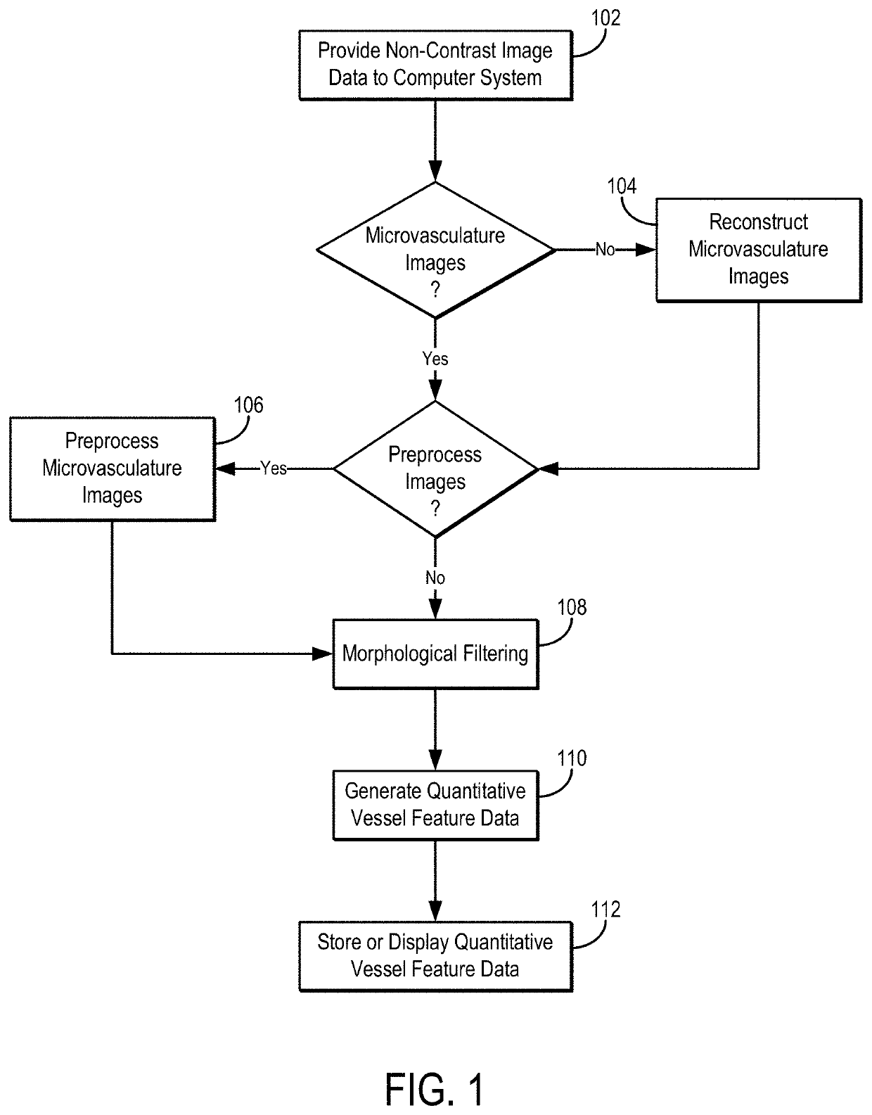
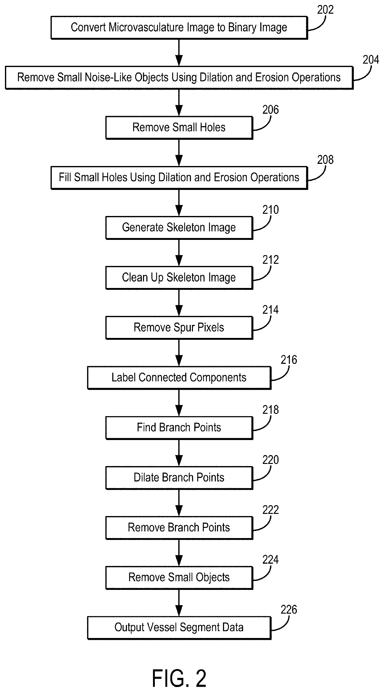
[0021]Described here are systems and methods for quantifying vessel features in non-contrast microvessel images obtained with an ultrasound imaging system. Vessel features that are quantified include, but are not limited to, vessel morphology features such as spatial vascularity pattern, bifurcation angle, Murray's deviation, fractal dimension, and closet vessel distance.

[0022]Advantageously, the present disclosure describes systems and methods for generating quantitative ultrasound biomarkers of microvessel morphology that correlate with the state of a disease in question. As one example, quantitative features related to the morphology and distribution of the vascular networks can be automatically extracted, estimated, or otherwise derived from ultrasound microvessel images reconstructed from contrast-free ultrasound data.

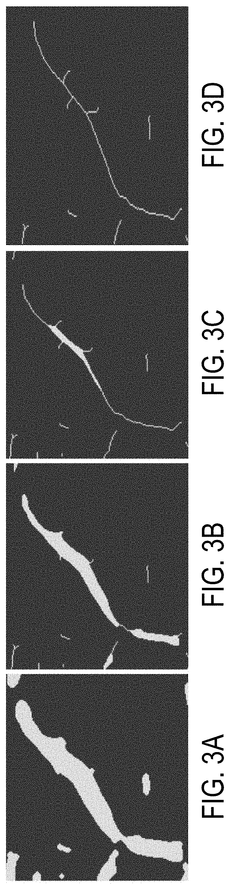
[0023]The systems and methods described in the present disclosure may be used to analyze the structure of blood vessels and to extract quantitative features from ...


PUM


Abstract

The present disclosure addresses a quantitative analysis of tumor vascularity patterns, with the goal of identifying biomarkers correlated with malignancy. Herein, we identify new types of quantitative ultrasound biomarkers of microvessel morphology that correlate with the state of the disease in question. We propose a novel method to automatically extract quantitative features related to the morphology and distribution of the vascular networks reconstructed from contrast-free ultrasound data. For instance, spatial vascularity pattern, bifurcation angle, Murray's deviation, fractal dimension and closet vessel distance were clearly depicted.

Description

CROSS-REFERENCE TO RELATED APPLICATIONS[0001]This application claims the benefit of U.S. Provisional Patent Application Ser. No. 63 / 074,085, filed on Sep. 3, 2020, and entitled “METHODS FOR EXTRACTING DIAGNOSTIC BIOMARKERS FROM ULTRASOUND MICROVESSEL IMAGES,” which is herein incorporated by reference in its entirety.STATEMENT REGARDING FEDERALLY SPONSORED RESEARCH[0002]This invention was made with government support under CA239548 awarded by the National Institutes of Health. The government has certain rights in the invention.BACKGROUND[0003]Angiogenesis, the growth of new microvessels, plays an essential role in pathogenesis and outcome of the deadliest maladies of the modern world, such as cancers. Quantification of microvessel morphological features could play an important role in disease diagnosis and tumor classification. For example, malignant tumor vascularity is known to present heterogeneous organization, structurally abnormal and chaotic. It has also been shown that microv...

Claims


Application Information

Patent Timeline
Patent Type & Authority Applications(United States)
IPC IPC(8): G06T7/00G06T7/155G06T7/73G16H50/30G16H50/20
CPCG06T7/0012G06T7/155G06T7/73G16H50/30G16H50/20G06T2207/30068G06T2207/30101G06T2207/30096G06T2207/30172G06T2207/20024G06T2207/10132G16H30/40G06T7/11G06T7/62
Inventor ALIZAD, AZRAFATEMI, MOSTAFATERNIFI, REDOUANEWANG, YINONG
Owner MAYO FOUND FOR MEDICAL EDUCATION & RES
Why Eureka
Try Eureka