
Anonymisation of medical patient images using an atlas

an atlas and medical patient technology, applied in the field of anonymisation of medical patient images, can solve problems such as hammering the usability of the disclosed method for radiotherapy planning

Active Publication Date: 2021-02-04
BRAINLAB
View PDF0 Cites 2 Cited by
  • Summary
  • Abstract
  • Description
  • Claims
  • Application Information

AI Technical Summary

Benefits of technology

The patent describes a method for creating medical images of a patient's body part for the purpose of radio therapy planning. The method involves first gathering information about the structure of the tissue in the image, by using an imaging modality like CT or MRI. The tissue is then segmented out, which is the process of separating the image data into different parts based on their appearance. This helps to create a more accurate and reliable image for use in planning the treatment. The method also helps to avoid distortion of the image due to bone, which can negatively impact the treatment's effectiveness.

Problems solved by technology

This avoids distortion of image information describing bony tissue which, if the disclosed method is used for radiotherapy planning, would severely hamper the usability of the disclosed method for radiotherapy planning because of the strong influence of bony tissue on the propagation of the treatment beam.

Method used


Image

  • Anonymisation of medical patient images using an atlas
  • Anonymisation of medical patient images using an atlas
  • Anonymisation of medical patient images using an atlas

Examples


Embodiment Construction

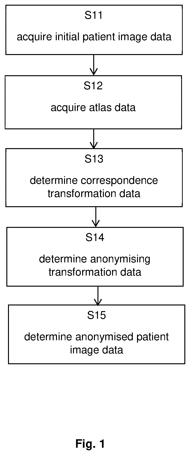
[0053]FIG. 1 illustrates the basic steps of the method according to the first aspect, in which step S11 encompasses acquisition of the initial patient image data, followed by step S12 which encompasses acquisition of the atlas data. Subsequent step S13 encompasses determination of the correspondence mapping data, which serves as a basis for determining in step S14 the anonymising transformation data. On the basis of the data determined and acquired in the foregoing steps, the anonymised patient image data is then determined in step S15.

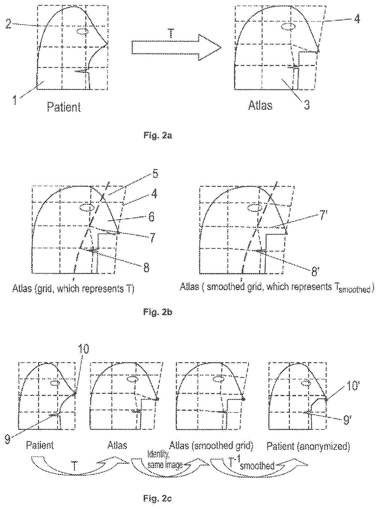
[0054]FIG. 2a describes the correspondence mapping T from a patient image showing a patient's head 1 onto an atlas showing an image-based model 3 of the head. The positions in the patient image are defined on a grid 2, and the positions in the atlas are defined in a grid 4.

[0055]FIG. 2b shows how an area 5 (delineated by a dashed line) in the image-based model 3 which shall serve as a region available for anonymising the medical image showing the pati...


PUM


Abstract

Disclosed is a computer-implemented method for generating an anonymized medical image of an anatomical body part of a patient, a corresponding computer program, a program storage medium storing such a program and a computer for executing the program, as well as a medical system comprising an electronic data storage device and the aforementioned computer. The disclosed method encompasses establishing a mapping from a patient image onto an atlas, changing that mapping, and applying the inverse of the changed mapping to the atlas in order to transform image content from the atlas to the patient image in order to achieve a deformed and thereby anonymised appearance of the patient image.

Description

FIELD OF THE INVENTION[0001]The present invention relates to a computer-implemented method for generating an anonymized medical image of an anatomical body part of a patient, a corresponding computer program, a program storage medium storing such a program and a computer for executing the program, as well as a medical system comprising an electronic data storage device and the aforementioned computer.TECHNICAL BACKGROUND[0002]It is desirable to anonymize patient faces in medical images in a way that the resulting data set still looks like a real patient data set so that image processing algorithms designed for medical data can still be applied without restrictions to the anonymized scans.[0003]So far face anonymization is done by coarse, non-anatomical modifications of the image in the facial region (e.g. by cutting off the face completely or by strong smoothing of the image in this region to remove biometric details). The result of such modifications looks distinctly different from...

Claims


Application Information

Patent Timeline
IPC IPC(8): G06F21/62G16H30/40G06T5/00
CPCG06F21/6254G06T5/002G16H30/40G06T5/70
Inventor BLUMHOFER, ANDREASSCHMALER, JENS
Owner BRAINLAB
Learn More