
Apparatus and method for promoting angiogenesis in ischemic tissue

a technology which is applied in the field of angiogenesis and angiogenesis in ischemic tissue, can solve the problems of limb/digital gangrene, non-healing ulcers, and rest pain in the foot, and achieve the effect of improving uptake into the surrounding ischemic tissue and reducing washou

Inactive Publication Date: 2018-12-27
ROCHA SINGH KRISHNA +1
View PDF25 Cites 2 Cited by
  • Summary
  • Abstract
  • Description
  • Claims
  • Application Information

AI Technical Summary

Benefits of technology

The present invention provides a way to deliver biologics, such as stem cells, to a specific area of an occluded vessel in a patient's body. This method creates a space between the layers of the vessel where the biologics can be placed, which improves their uptake and reduces the risk of them being washed out or carried into the systemic circulation. The invention also reduces the amount of biologics needed for treatment and improves the viability of the cells. The delivery device used in this method has multiple small openings that are designed to minimize damage to the biologics and allow for a more uniform distribution. Overall, this method helps to promote the growth of new blood vessels and improve the function of the surrounding tissue.

Problems solved by technology

Critical limb ischemia (CLI) is the most advanced stage of PAD, and affects more than 500,000 people annually, causing rest pain in the foot, non-healing ulcers, delayed wound healing, limb / digital gangrene, and may eventually lead to amputation.
However, many patients with severe, diffuse arterial occlusive disease, as is typically seen in CLI, are either not ideal candidates for an endovascular approach to percutaneous revascularization due to their significant co-morbidities (i.e. renal dysfunction, myocardial dysfunction) or significant functional or nutritional debilitation.
Moreover, when arterial occlusions are extensive and severely calcified, which is the typical in CLI patients with femoropopliteal and infrapopliteal occlusive disease, interventional attempts to re-establish vessel patency to improve tissue perfusion are frequency sub-optimal or unsuccessful due to inability to traverse these complex, long arterial occlusions.
Given this technical failure to re-establish tissue perfusion, amputation is frequently required as a life saving measure, resulting in long-term disability, a diminished quality of life and substantial expenditures to the health care system.
These delivery methods have remained in use, and relatively static, for over a decade, and may significantly hamper the effectiveness of stem-cell therapy.
Specifically, stem cell viability and retention rates after conventional delivery methods are extremely low, typically less than 10% of the injected number.
However, simply injecting a larger number of stem cells cannot compensate for inefficient delivery modalities.
Furthermore, stem cells have relatively large diameters and may occlude vessels, compromising their therapeutic effect and potentially contributing to worsening ischemic symptoms.
First, stem cells differ from traditional therapeutics in that the cells are fragile and extremely sensitive to their microenvironments.
Previously known delivery methods deposit the stem cells directly at the occlusion sites, where—due to ischemia, hypoxia, oxidative stress, or inflammation—the microenvironment may be harsh, and contribute to massive cell apoptosis.
Further, the cell injection process may itself contribute to poor cell viability, as injection may cause mechanical disruption to the stem cells, e.g., barotrauma caused by fluid sheer and extreme pressure fluctuations.
Moreover, in certain applications, such as delivery of stem cells to long femoropopliteal occlusions, the migration of stem cells to ischemic areas may be impeded by the long, calcified occlusions.

Method used


Image

  • Apparatus and method for promoting angiogenesis in ischemic tissue
  • Apparatus and method for promoting angiogenesis in ischemic tissue
  • Apparatus and method for promoting angiogenesis in ischemic tissue

Examples


Embodiment Construction

[0043]Arteries and veins generally are comprised of three layers: the innermost layer called the intimal layer, the outermost layer called the adventitial layer, and the medial layer located in between the intimal and the adventitial layers. The intimal and medial layers are readily separated from the adventitial layer. For example, it is known that when attempting to pass an occlusion with a guidewire, the guidewire may sometimes inadvertently penetrate the subintimal space between the intimal and the adventitial layer. Hereinafter, in the context of this specification, the term “intimal layer” refers to the intima / media that adjoins the occluded vessel lumen, while the term “adventitial layer” refers to the outer layer of the vessel that may be separated from the intimal layer when a subintimal space is created.

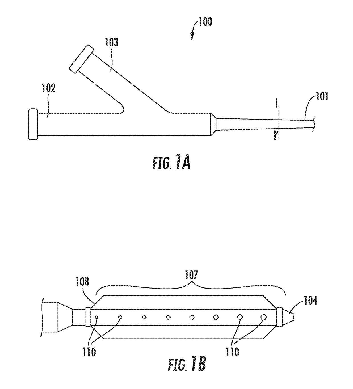
[0044]Referring to FIGS. 1A and 1B, a biologics delivery device constructed in accordance with the principles of the present invention is described. Biologics delivery devi...


PUM


Abstract

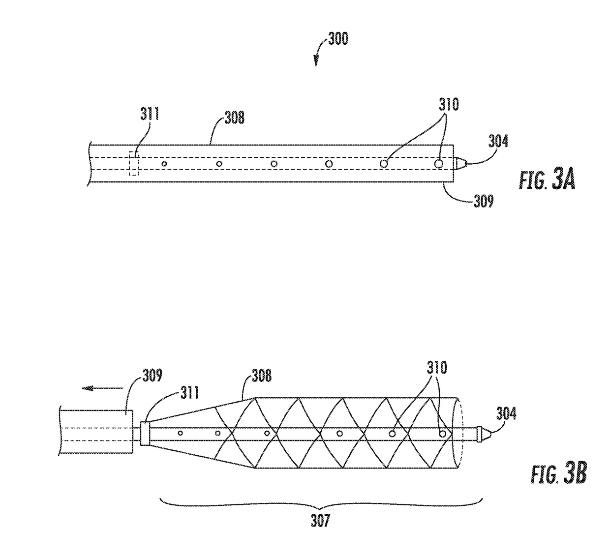
A biologics delivery device and method of use for promoting angiogenesis in occluded vessels and ischemic tissue of a patient are provided, wherein the biologics delivery device includes a catheter having a proximal end, a distal region having a distal end, and a side wall defining a catheter lumen; an expandable member disposed in the distal region, the expandable member configured to support a subintimal space in an occluded blood vessel of a patient, and to transition between a collapsed state and an expanded state; and a hollow needle having a penetration tip deployable from inside the catheter lumen to outside the catheter lumen, and into tissue surrounding the occluded blood vessel, wherein the expandable member in the expanded state allows the flow of oxygenated blood to the occluded blood vessel. Methods of using the inventive biologics delivery device also are provided to deposit the biologic from a subintimal space to tissue surrounding the occluded blood vessel.

Description

CROSS-REFERENCE TO RELATED APPLICATIONS[0001]This application is a continuation-in-part application of U.S. patent application Ser. No. 15 / 072,249, filed Mar. 16, 2016, the entire contents of which are incorporated herein by reference.FIELD OF THE INVENTION[0002]The present invention is directed to apparatus and methods for promoting tissue perfusion in the vascular territory served by vessels occluded by severe atherosclerosis by delivering biologics into a subintimal space, and / or by delivering biologics from the subintimal space into tissue surrounding the vessel.BACKGROUND OF THE INVENTION[0003]Peripheral arterial disease (PAD) is a highly prevalent disease affecting over 12 million people in the United States (Golomb et al., Circulation, 2006). As PAD progresses, atherosclerosis and chronic inflammation can result in markedly reduced blood flow to the legs, feet, and hands. Critical limb ischemia (CLI) is the most advanced stage of PAD, and affects more than 500,000 people annu...

Claims


Application Information

Patent Timeline
Patent Type & Authority Applications(United States)
IPC IPC(8): A61M25/00A61M25/04A61M25/01
CPCA61M2025/0197A61M25/04A61M25/007A61M25/0074A61M2025/0092A61M25/0084A61M2205/32
Inventor ROCHA-SINGH, KRISHNAMARTINEZ-SINGH, KRISHNA
Owner ROCHA SINGH KRISHNA
Learn More