Methods for isolating microvesicles
- Summary
- Abstract
- Description
- Claims
- Application Information
AI Technical Summary
Benefits of technology
Problems solved by technology
Method used
Image


Examples
example 1
Plasma Microvesicular RNA can be Isolated Using a 0.65 um Positively Charged Q PES Filter in a Vacuum Format
[0244]Normal control plasma was obtained from Bioreclamation LLC. Blood was collected into K2 EDTA tubes and mixed well with complete inversions. The tubes were centrifuged at 1300×g for 10 min to separate the blood cells and plasma. Then, the plasma was filtered through a 0.8 μm mixed cellulose ester filter (Millipore, Billerica, Ma., USA) to remove cell debris and platelets, and divided into 1 mL aliquots. Aliquots were frozen at −80° C. until needed.
[0245]Isolation of 4 mL plasma microvesicular RNA was conducted using ultracentrifugation or Fast Trap Adenovirus purification and concentration kit (Millipore, Billerica, Ma., USA) 0.65 um positively charged Q polyethersulfone vacuum filtration.
[0246]In the ultracentrifugation method, one 1 mL aliquots of plasma from four subjects were transferred to a 5 mL polyallomer tube (Beckman-Coulter, Miami, Fl., USA) containing 8 μL RNa...
example 2
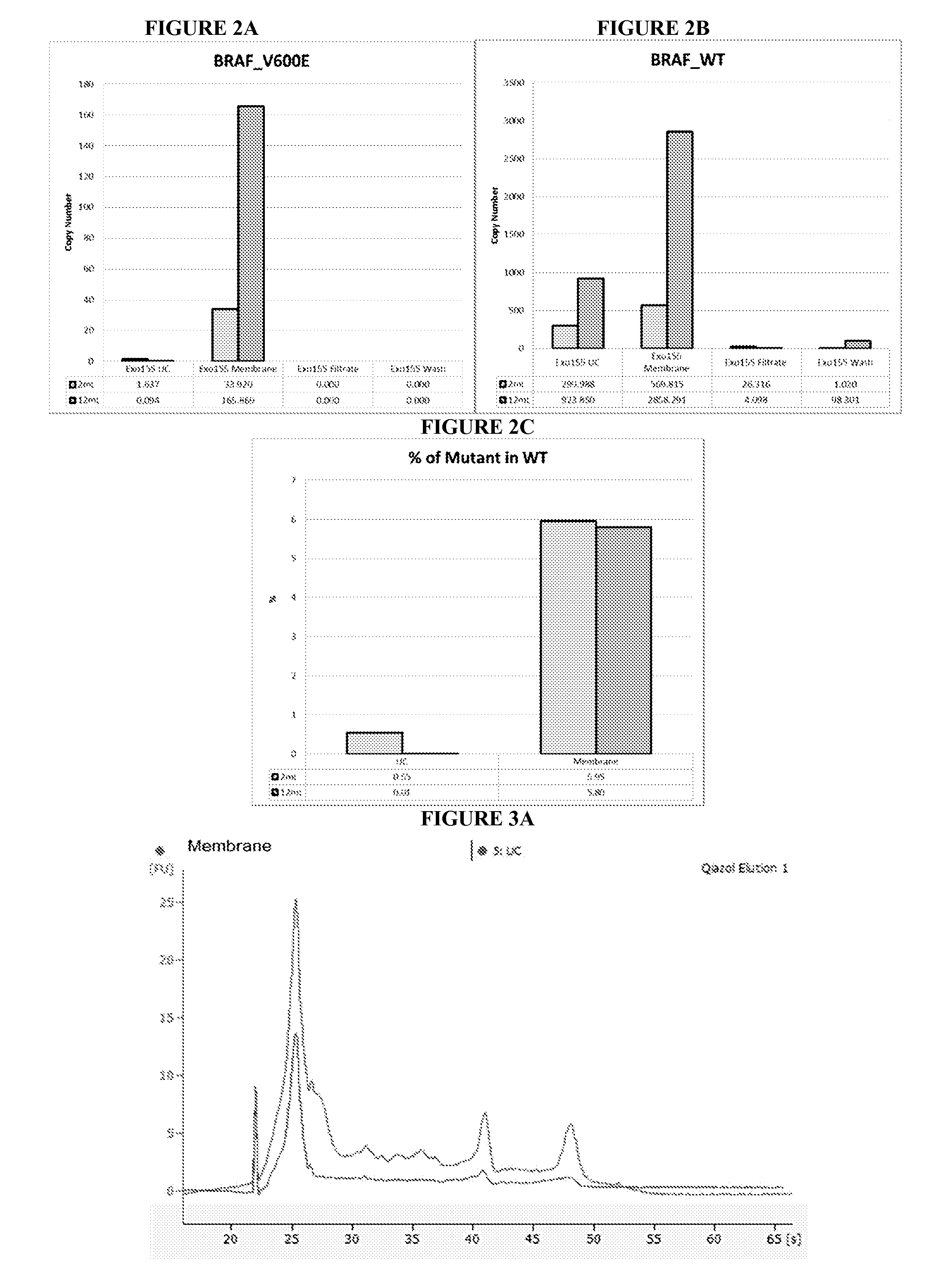
Detecting BRAF_V600E Mutation in Plasma Sample from Melanoma Patient Using Millipore Fast Trap Virus Purification Column
[0251]Melanoma plasma was obtained from Ludwig Maximilians University in accordance with protocols approved by the IRB and as described in EXAMPLE 1.
[0252]Isolation of 2 mL and 12 mL plasma microvesicular RNA was conducted using ultracentrifugation or Fast Trap Adenovirus purification and concentration kit (Millipore, Billerica, Ma., USA) 0.65 um positively charged Q polyethersulfone vacuum filtration.
[0253]In the ultracentrifugation method, 2 mL and 3×4 mL plasma were transferred to a 5 mL polyallomer tube (Beckman-Coulter, Miami, Fl., USA) containing 8 μL RNasin Plus (40 U / μl, Promega, Madison, Wi., USA) RNase inhibitor, and incubated for 5 minutes at room temp. Following incubation, the 2 mL and each 4 mL plasma was diluted in 3 mL and 1 mL PBS, respectively. Microvesicles were pelleted and lysed as described in EXAMPLE 1.
[0254]In the 0.65 um positively charged ...
example 3
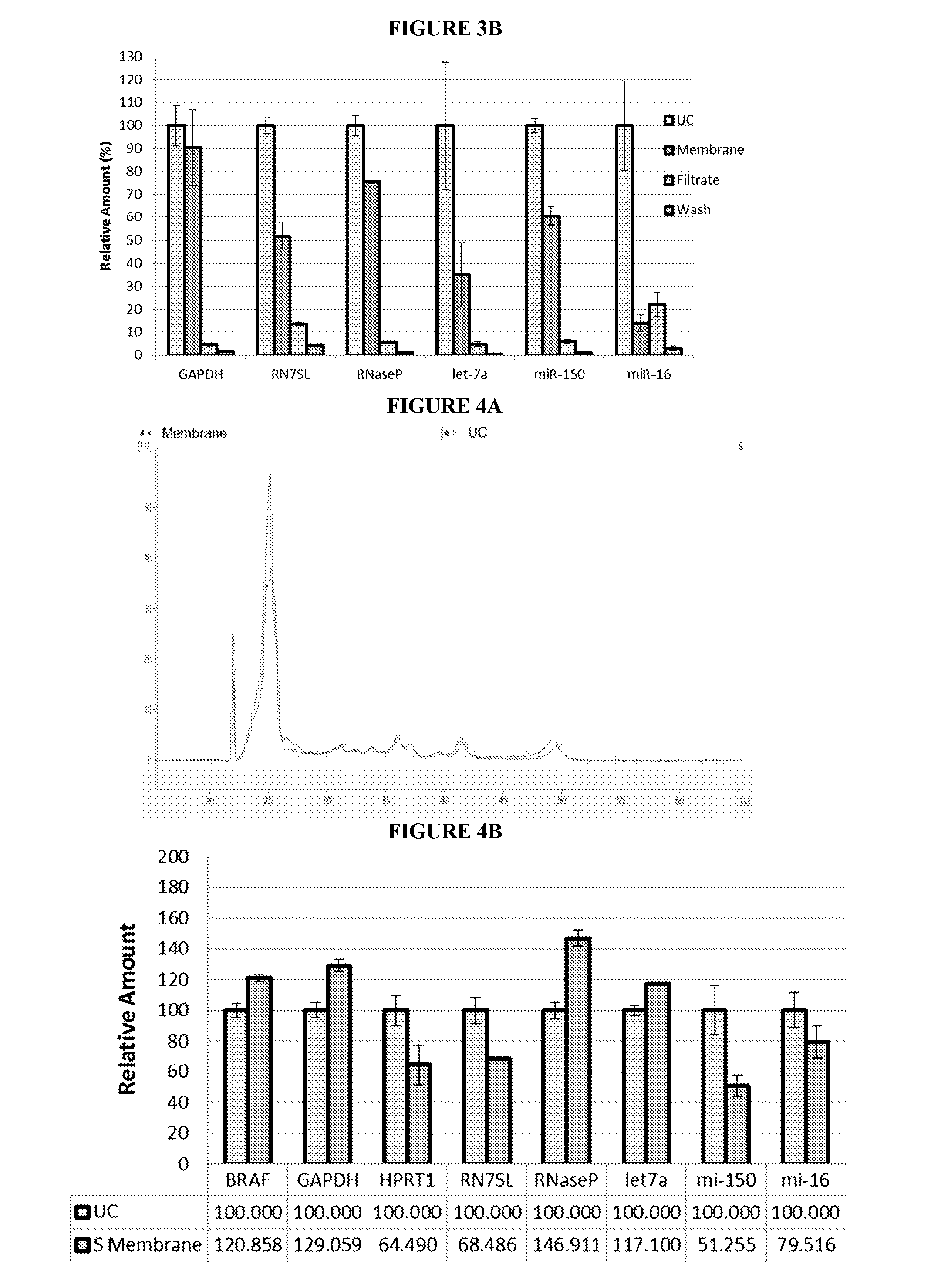
Regenerated Cellulose Spin Column
[0259]Normal control plasma was obtained from Bioreclamation LLC, as described in EXAMPLE 1.
[0260]Isolation of 4 mL plasma microvesicular RNA was conducted using ultracentrifugation or 3 um positively charged Q regenerated cellulose centrifugal filtration.
[0261]The ultracentrifugation sample was prepared, pelleted, and lysed as described in EXAMPLE 1.
[0262]In the 3 um positively charged Q regenerated cellulose centrifugal filtration method, 1 mL aliquots of plasma from the same four subjects used in the ultracentrifugation method were pooled and diluted with 444 ul 10× Loading Buffer (Sartorius). Before the plasma sample was applied, the filter was conditioned by passing through 5 mL 1× Washing Buffer (Sartorius) at 500×g for 5 min. Then, the plasma sample was passed through the filter at 500×g for 5 min. The filtrate was saved for further analysis. The filter was washed twice with 18 mL Washing Buffer (Sartorius) at 500×g for 5 min. The first wash w...
PUM
| Property | Measurement | Unit |
|---|---|---|
| Pore size | aaaaa | aaaaa |
| Fraction | aaaaa | aaaaa |
| Electric charge | aaaaa | aaaaa |
Abstract
Description
Claims
Application Information
 - R&D
- Intellectual Property
- Life Sciences
- Materials
- Tech Scout
- Unparalleled Data Quality
- Higher Quality Content
- 60% Fewer Hallucinations
Browse by: Latest US Patents, China's latest patents, Technical Efficacy Thesaurus, Application Domain, Technology Topic, Popular Technical Reports.
© 2025 PatSnap. All rights reserved.Legal|Privacy policy|Modern Slavery Act Transparency Statement|Sitemap|About US| Contact US: help@patsnap.com