
Marker for detecting myogenic disease and detection method using the same

a technology of myogenic disease and detection method, which is applied in the field of myogenic disease detection markers, can solve the problems of disadvantageously misdiagnosis, high creatine kinase level in normal individuals, and leakage of enzymes including creatine kinase into blood, and achieve the effect of low invasiveness

Inactive Publication Date: 2015-07-30
INTPROP STRATEGY NETWORK INC
View PDF1 Cites 1 Cited by
  • Summary
  • Abstract
  • Description
  • Claims
  • Application Information

AI Technical Summary

Benefits of technology

[0018]According to a marker for detecting myogenic disease of the present invention, there can be provided a marker for detecting myogenic disease affecting a test subject without being influenced by exercise stress.
[0019]According to a method for detecting myogenic disease of the present invention, there can be provided a method capable of detecting the presence or absence of myogenic disease affecting a test subject, wherein the method is low invasive to the test subject and is insusceptible to exercise stress to the test subject.

Problems solved by technology

However, the enzymes including creatine kinase also leak into blood by myocyte necrosis attributed to exercise stress.
As a result, even normal individuals exhibit a high level of creatine kinase or the like and are disadvantageously misdiagnosed.
For accurate diagnosis, test subjects must be placed in the resting state before blood collection, and this process is burdensome.

Method used


Image

  • Marker for detecting myogenic disease and detection method using the same
  • Marker for detecting myogenic disease and detection method using the same
  • Marker for detecting myogenic disease and detection method using the same

Examples


example 1

Verification of Marker for Detecting Muscular Dystrophy Using Mouse Muscular Dystrophy Models

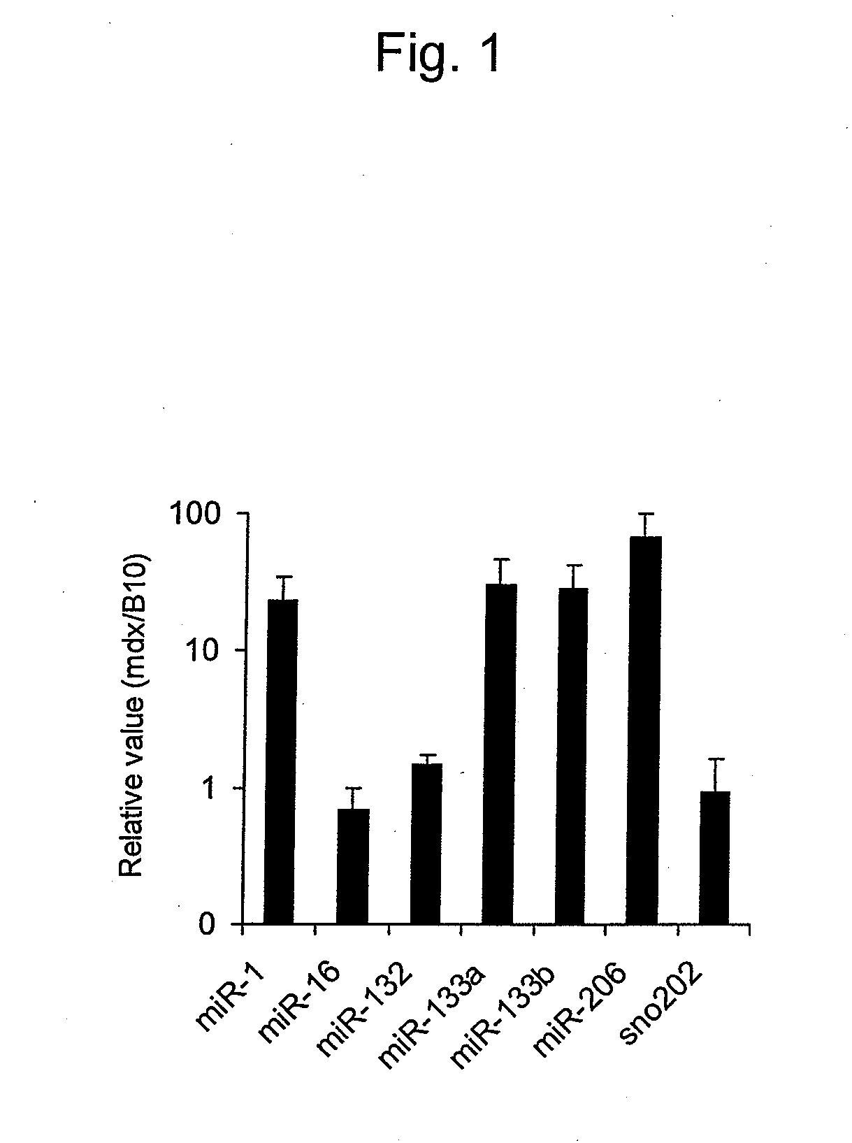
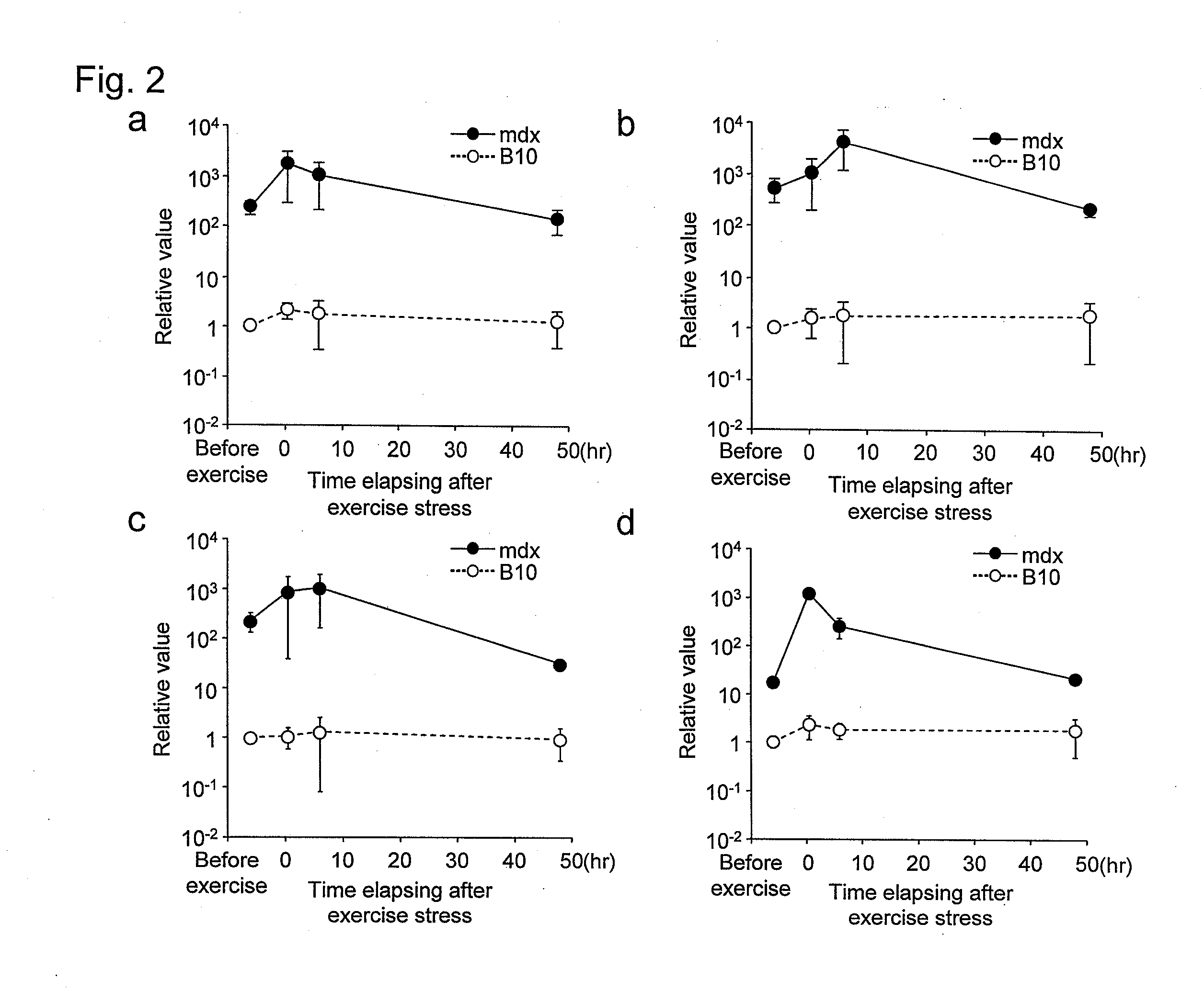
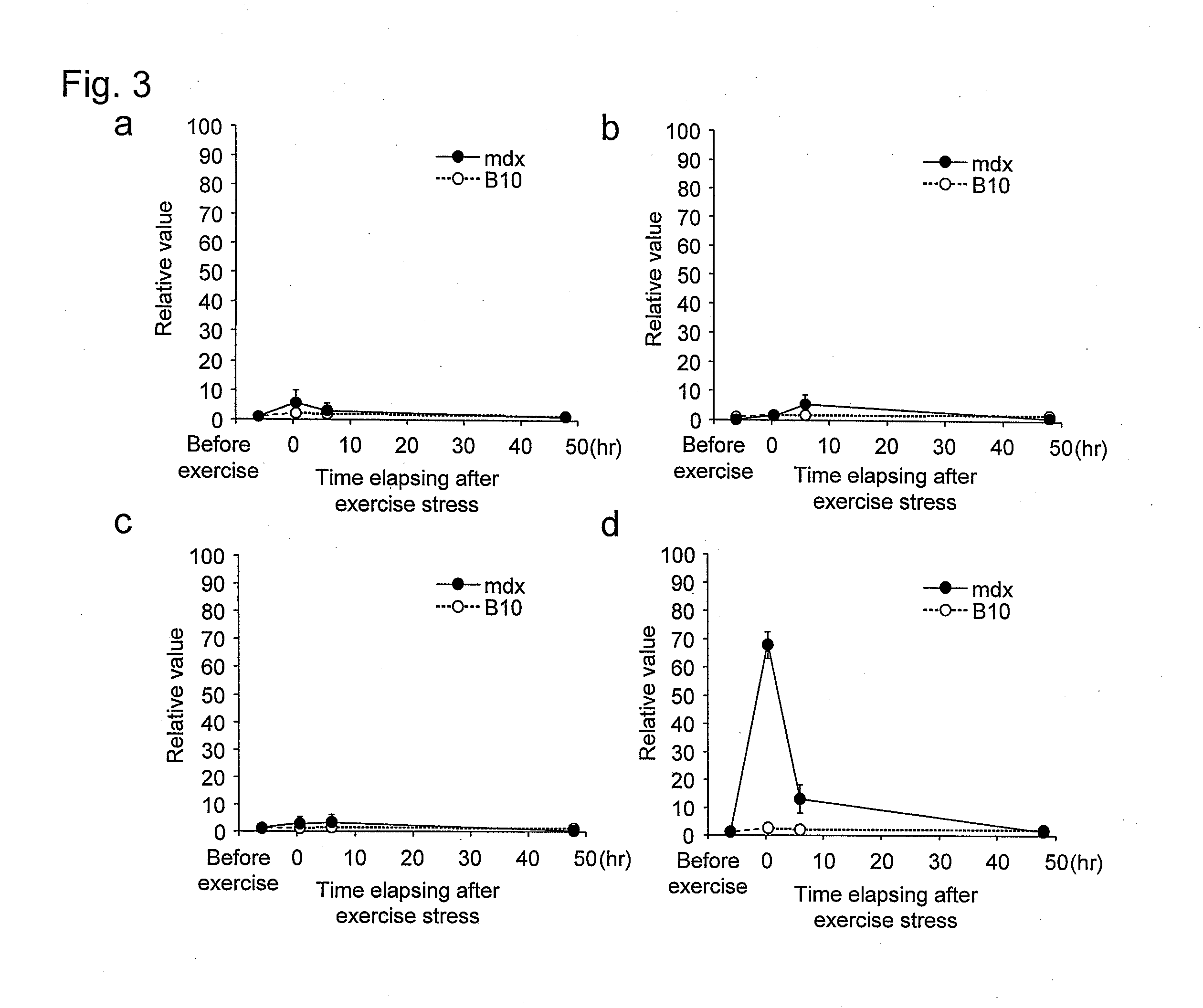
[0080]The effects of the marker for detecting muscular dystrophy of the present invention and the method for detecting muscular dystrophy using the same were validated using mouse muscular dystrophy models.

(Materials)

[0081]The mouse muscular dystrophy models used were mdx mice (mdx / B10) (male individuals; 8 weeks old), disease models of Duchenne muscular dystrophy. Duchenne muscular dystrophy is developed by the deficiency of the dystrophin gene caused by X-linked recessive inheritance. Also, B10 mice (male individuals; 8 weeks old) were used as a control (normal individual) group. In this context, the mdx mice have the same genetic background as in the B10 mice except for the deficiency of the dystrophin gene.

(Method)

[0082]Blood Collection and Preparation of Serum

[0083]Each of the mice was brought in an animal experiment facility and then separately caged and preliminarily raised for 1 week...

example 2

Verification of Marker for Detecting Muscular Dystrophy Using Dog Muscular Dystrophy Models

[0105]The effects of the marker for detecting muscular dystrophy of the present invention and the detection method using the same were validated using dog muscular dystrophy models. Although mdx mice, unlike human muscular dystrophy patients, do not show symptoms such as gait abnormality, muscular dystrophy dogs deficient in the dystrophin gene by X-linked recessive inheritance as in the mdx mice show symptoms such as gait abnormality similar to those in humans. Thus, effects more similar to those seen in humans can be verified.

(Materials)

[0106]The dog muscular dystrophy models used were beagles of CXMDJ lineage having abnormal dystrophin genes on the X-chromosomes, carrier dogs thereof (female beagles having abnormality in one dystrophin gene on the X chromosomal pair), and normal dogs (beagles free from such abnormality in the dystrophin gene).

(Method)

[0107]100 μL or more of blood was collec...


PUM

PropertyMeasurementUnit
volumeaaaaaaaaaa
volumeaaaaaaaaaa
volumeaaaaaaaaaa

Abstract

It is intended to provide a low invasive and highly sensitive method for detecting myogenic disease without being influenced by exercise stress. The amount of any one or more of miR-1, miR-133a, miR-133b, and miR-206 in the blood of a test subject is measured. When the amount of the miRNA in the blood is statistically significantly higher than that of corresponding miRNA in the blood of a normal individual, it indicates that the test subject is suffering from myogenic disease.

Description

TECHNICAL FIELD[0001]The present invention relates to a marker for detecting myogenic disease and a method for detecting the presence or absence of myogenic disease, particularly, muscular dystrophy, affecting a test subject using the same.BACKGROUND ART[0002]Muscular dystrophy, a type of myogenic disease, is a progressive genetic disease that causes muscle wasting or weakness resulting from the degeneration or necrosis of muscle fibers in skeletal muscles. This disease is known to have various types, such as Duchenne, Becker, limb-girdle, and facioscapulohumeral types, depending on the mode of inheritance or clinical conditions (Non Patent Literature 1).[0003]Muscular dystrophy is comprehensively diagnosed by means of clinical conditions, blood test, examination findings, electromyography, muscle biopsy, genetic test, etc. Of them, the blood test is conducted by the determination of the amount of an enzyme such as creatine kinase, lactate dehydrogenase, glutamic oxaloacetic transam...

Claims


Application Information

Patent Timeline
Patent Type & Authority Applications(United States)
IPC IPC(8): C12Q1/68
CPCC12Q2600/178C12Q1/6883C12N2310/141C12N15/111C12N2320/10C12Q2600/158
Inventor HASHIDO, KAZUOMIZUNO, HIDEYA
Owner INTPROP STRATEGY NETWORK INC
Learn More