Composition for diagnosis of lung cancer and diagnosis kit of lung cancer

a lung cancer and composition technology, applied in the field of composition for diagnosis of lung cancer and a lung cancer diagnosis kit, can solve the problems of lack of a high-sensitivity early diagnosis method, lack of commercialized biomarkers capable of specifically diagnosing lung cancer, and insufficient specificity and sensitivity. achieve excellent sensitivity and specificity

Inactive Publication Date: 2014-09-04
SEOUL NAT UNIV R&DB FOUND
View PDF1 Cites 1 Cited by
  • Summary
  • Abstract
  • Description
  • Claims
  • Application Information

AI Technical Summary

Benefits of technology

[0011]In the present invention, through direct discovery of a lung cancer-specific biomarker from a tissue, lung cancer can be quickly, simply, and accurately diagnosed.
[0019]The inventive composition for diagnosis of lung cancer is excellent in sensitivity and specificity, and thus can be usefully used in early diagnosis of lung cancer.

Problems solved by technology

Such a tendency is caused by limited subjective symptoms, and the absence of an early diagnosis method having a high sensitivity.
However, they have not yet sufficiently shown specificity and sensitivity.
Thus, in various diseases, cross-reactivity exists, while there is no method for complementing this.
However, up to now, there is no commercialized biomarker capable of specifically diagnosing lung cancer.
Thus, it is difficult to find a tissue-specific origin protein, and also there exist many difficulties in detection due to a wide dynamic range of a blood protein.

Method used


Image

  • Composition for diagnosis of lung cancer and diagnosis kit of lung cancer
  • Composition for diagnosis of lung cancer and diagnosis kit of lung cancer
  • Composition for diagnosis of lung cancer and diagnosis kit of lung cancer

Examples


example 1

Test Sample Collection

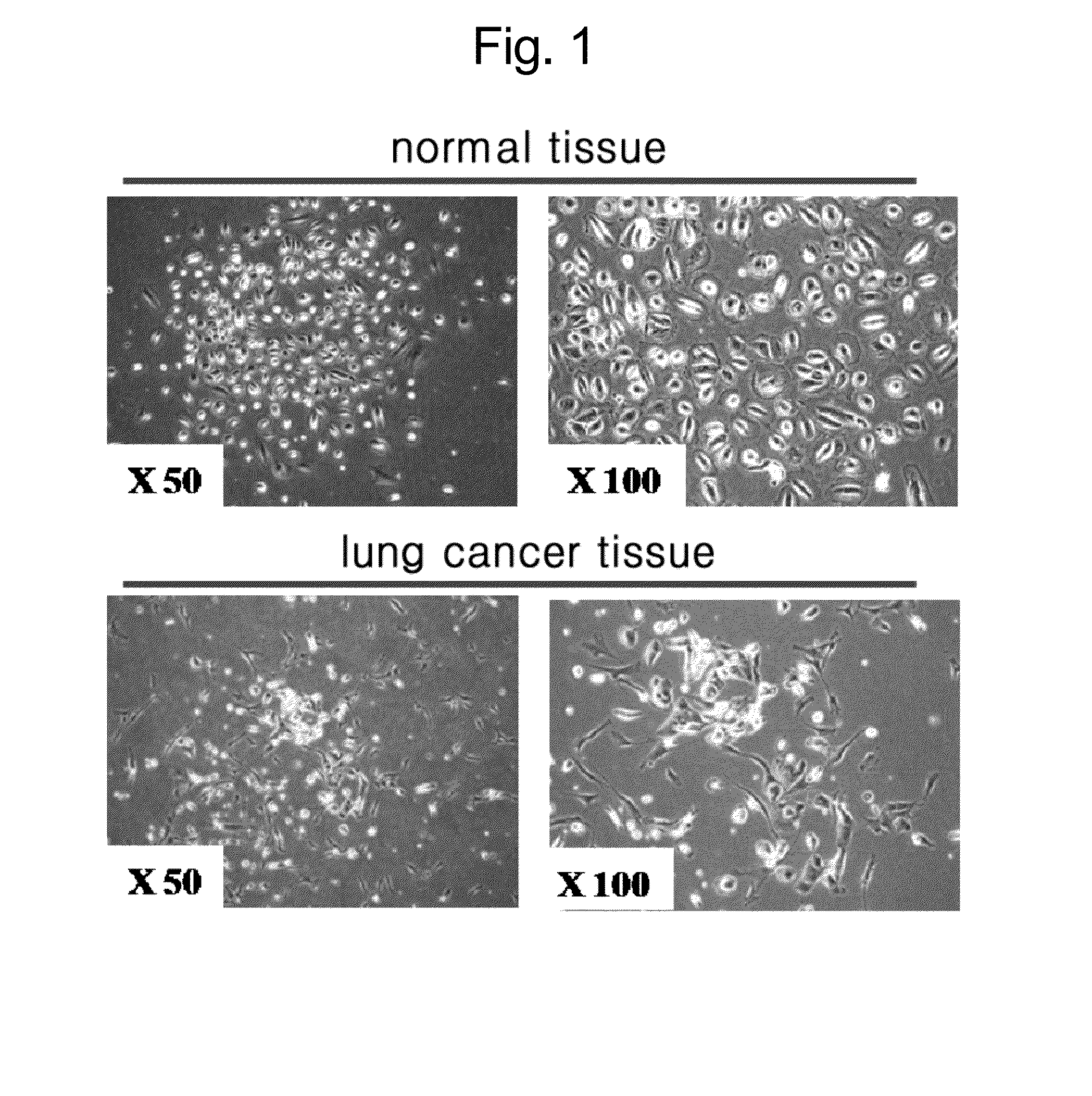
[0053]A system for primary culture using a lung cancer tissue and a surrounding normal tissue obtained from a lung cancer patient was secured. The lung cancer tissue and the surrounding normal tissue were maintained in a fresh state, and were chopped by using scissors or a blade in accordance with a mechanical method. Then, they were treated with collagenase type I, reacted for 30 minutes to 90 minutes at 37° C. so as to separate single cells. Then, they were plated onto a medium containing 10% FBS, and cultured.

[0054]In a case of a cell separated from the normal tissue, a typical epithelial cell type of pebble-shaped circular cells were grown, while in a case of a cell separated from the lung cancer tissue, fibrocyte-like shaped cells were grown in a long form (see FIG. 1).

example 2

Separation and Concentration of a Secreted Protein

[0055]Cells from Example 1 were subcultured twice to three times so as to collect a protein secreted during a culturing process. In subculture 3, the same number of cells were counted, and they were transferred to a new culturing plate, and cultured for 48 hours to collect a medium including a secreted protein.

[0056]Then, in order to remove the FBS content contained in a subculture medium of the normal cell plate and the lung cancer cell plate, each plate was sufficiently washed. Then, the medium was replaced by a serum free media, and culturing was performed for 24 to 48 hours. In this process, in order to obtain a protein secreted from each cell, a TCA precipitation method was used for protein concentration.

example 3

Separation and Coomassie Blue Staining of SDS-PAGE

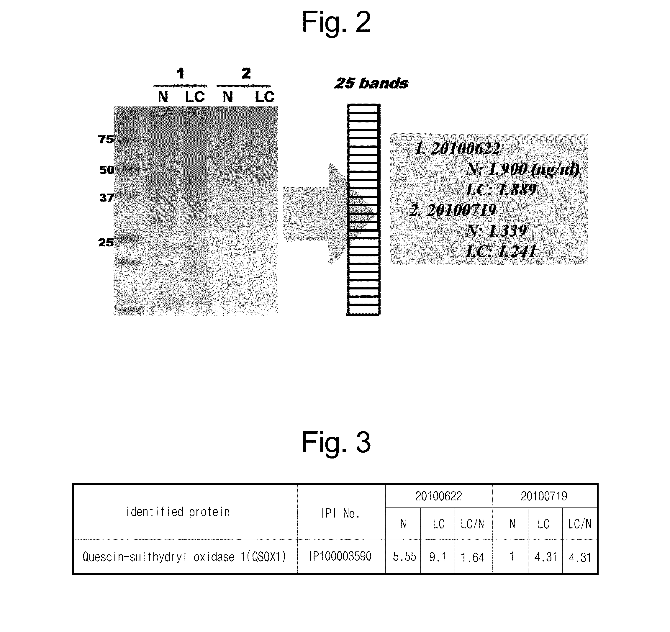
[0057]The protein from Example 2, which was secreted from the normal tissue cell and the lung cancer tissue cell and concentrated, was separated through SDS-PAGE, and the gel was washed with ddH2O three times (each for 5 minutes). Then, the protein was added with Coomassie G250 Stain (BioRad, Hercules, Calif.), and softly shaken at room temperature for 1 hour for staining. In the coomassie staining, in each pair (N: a protein secreted from a normal tissue, LC: a protein secreted from a lung cancer tissue), the proteins were used for the experiment in the same amount (see FIG. 2).


PUM

PropertyMeasurementUnit
electrophoresisaaaaaaaaaa
fluorescence activated cell sorteraaaaaaaaaa
CTaaaaaaaaaa

Abstract

Disclosed is as a biomarker useful in early diagnosis of lung cancer, at least one protein selected from the group including Quescin-sulfhydryl oxidase 1, Fibrillin-1, Isoform A of Lamin-A / C, Latent-transforming growth factor beta-binding protein 2, Galectin-1, highly similar to Dickkopf-related protein 3, Isoform A1-B of Heterogeneous nuclear ribonucleoprotein A1, 14-3-3 protein epsilon, Stanniocalcin-2, Cystatin-C, Isoform 1 of Connective tissue growth factor, Profilin-1, Isoform 1 of Extracellular matrix protein 1, Histone H2B type 2-E, Kinesin-like protein KIF26A, Zinc finger protein 516, and Isoform 1 of A-kinase anchor protein 9.

Description

CROSS REFERENCE TO RELATED APPLICATIONS[0001]This application is a divisional of U.S. Ser. No. 13 / 351,631, filed Jan. 17, 2012, which claims priority to and the benefit of Korean Application No. 10-2011-0104949 filed on Oct. 14, 2011, in the Korean Patent Office, the entire contents of which are incorporated herein by reference.BACKGROUND OF THE INVENTION[0002]1. Field of the invention[0003]The present invention relates to a composition for diagnosis of lung cancer, and a lung cancer diagnosis method using the same, in which the composition includes a lung cancer-specific protein marker whose expression is increased or decreased in a lung cancer tissue.[0004]2. Description of the Prior Art[0005]Lung cancer is currently one of the cancers showing high lethality overseas as well as domestically. Such a tendency is caused by limited subjective symptoms, and the absence of an early diagnosis method having a high sensitivity. At present, the diagnosis of lung cancer relatively highly dep...

Claims


Application Information

Patent Timeline
Patent Type & Authority Applications(United States)
IPC IPC(8): G01N33/573G01N33/574
CPCG01N33/57423G01N33/573C12Q1/6886G01N33/5302G01N33/6857G01N33/6893
Inventor CHO, JE YOELSUNG, HYE JIN
Owner SEOUL NAT UNIV R&DB FOUND
Why Eureka
Try Eureka