Detection of Protein Translocation by Beta-Galactosidase Reporter Fragment Complementation

a reporter fragment and protein technology, applied in the field of molecular biology, can solve the problems of difficult control of stoichiometry and the location of dye attachment, poorly understood mechanisms involved in nuclear translocation and targeting of steroid receptors to regulatory sites in chromatin, and inability to track these events. , to achieve the effect of precise and accurate monitoring

Active Publication Date: 2012-03-29
THE BOARD OF TRUSTEES OF THE LELAND STANFORD JUNIOR UNIV
View PDF4 Cites 1 Cited by
  • Summary
  • Abstract
  • Description
  • Claims
  • Application Information

AI Technical Summary

Benefits of technology

[0015]Precise and accurate monitoring of protein translocation permits screening for and understanding the interplay key components of biologically relevant cellular signaling pathways. Moreover, such assays provide a means for screening and identifying modulators of protein translocation events that may be useful in the diagnosis, treatment, or prevention of disorders and diseases that can be impacted through protein translocation events. Enzymatic assays confer several advantages in monitoring protein translocation events including signal amplification and a wide variety of substrates for in vivo and in vitro detection. Provided herein is a novel assay system for monitoring protein translocation based on enzymatic complementation.

Problems solved by technology

However, the mechanisms involved in nuclear translocation and targeting of steroid receptors to regulatory sites in chromatin have been poorly understood.
Current technologies to track these events are limited to biochemical fractionation or fusion to fluorescent proteins.
However, the stoichiometry and locations of dye attachment are often difficult to control, and careful repurification of the proteins is usually necessary.
Biochemical methods are often the most sensitive and quantitative however they are limited by their ability to discern sub-cellular structures without contamination from other organelles.
In addition, the number of manipulations involved in preparing the samples makes these methods cumbersome and prone to high variability.
However the large amounts of protein necessary for efficient imaging make these experiments difficult to perform with toxic proteins and the supra-physiological levels of target protein can affect the quality of the data obtained.
However the difficulties associated with quantification of these events, such as small increases in fluorescence, high cell to cell variability, and the necessity for high expression levels of the fusion protein, prohibit its use in certain applications and limit the data to mainly qualitative measurements.

Method used


Image

  • Detection of Protein Translocation by Beta-Galactosidase Reporter Fragment Complementation
  • Detection of Protein Translocation by Beta-Galactosidase Reporter Fragment Complementation
  • Detection of Protein Translocation by Beta-Galactosidase Reporter Fragment Complementation

Examples


example 1

Design of the Minimal Fragment

[0145]α-complementation of β-galactosidase involves a deletion mutant, the ω fragment (ω), that is missing amino acids 11-41. In E. coli and in vitro, it is possible to complement this mutant in trans by providing the α peptide which encodes amino acids 3-41. However, previous studies in mammalian cells reported that the smallest complementing fragment contained amino acids 1-71. Because a smaller peptide enhances the efficiency of anal sensitivity, the smallest α peptide(s) that would complement robustly in mammalian cells when fused to an exogenous protein at the N or C terminus was determined. The myoblast cell line C2C12 was engineered to stably express the co fragment by retroviral transduction. The α potion of β-galactosidase was fused to the C-terminus of GFP and deletions were made stating at amino acids 137 and continuing through amino acids 38. The products were transduced into the parental cell line expressing the ω-fragment, and the cells we...

example 2

Nuclear Translocation Assay

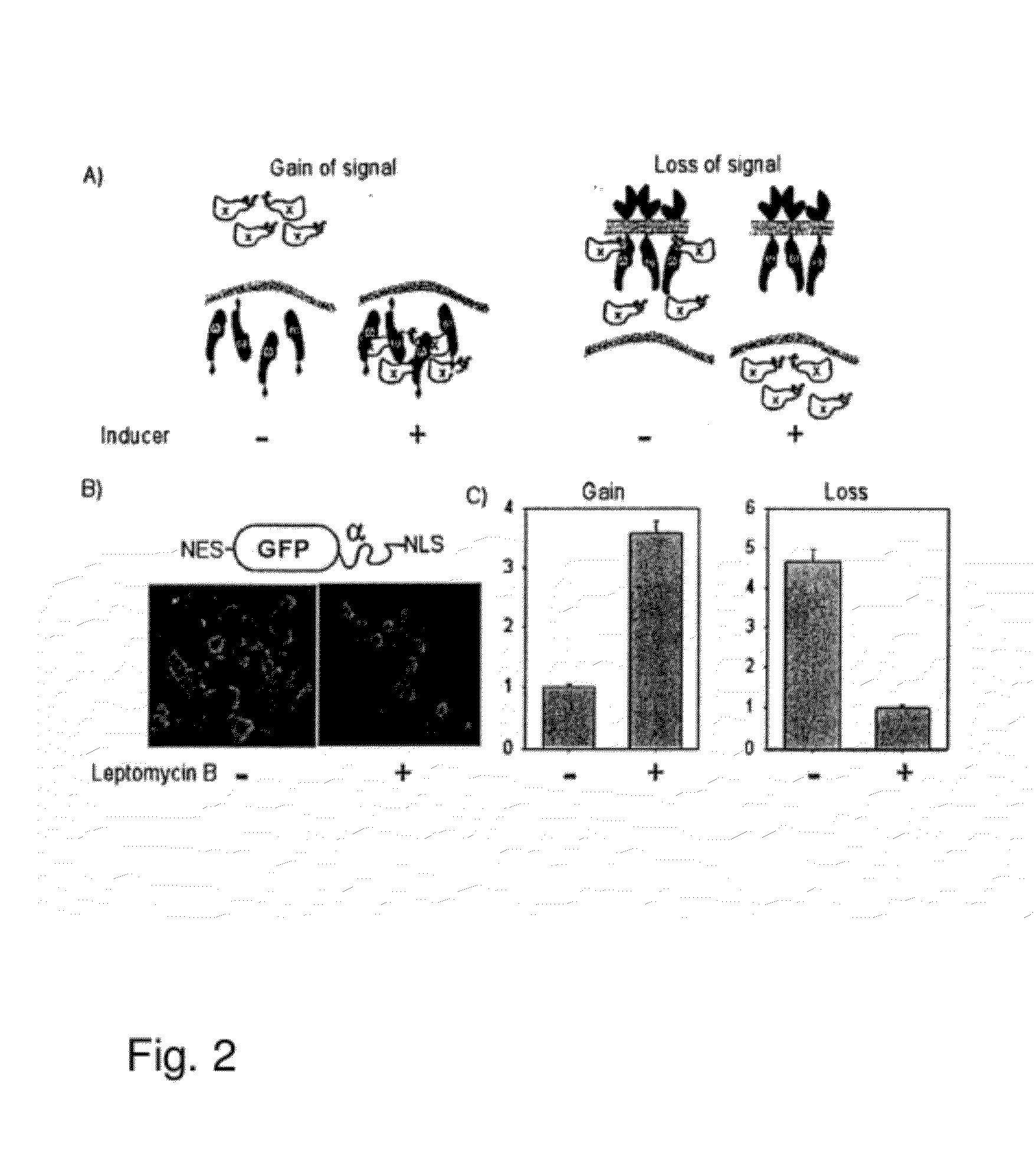
[0148]In order to utilize a complementation as a method of quantitatively assessing nuclear translocation events, two systems were designed that are distinct in the localization of the ω-fragment. FIG. 2A is a schematic showing the design of the nuclear translocation assay. In the let panel, the ω fragment is localized to the nucleus with a nuclear localization signal (NLS) and the cytosolic protein of interest is fused to the minimal α peptide. Upon stimulation the α fusion moves to the nucleus and complements the co, increasing β-galactosidase activity. In the right panel, the co fragment is tethered to the plasma membrane using the extracellular and transmembrane regions of EGFR. The cytosolic α-fusion complements spontaneously until stimulation when it translocates to the nucleus which results in a loss of enzyme activity.

[0149]The gain of signal assay (FIG. 2A left panel) localized the ω to the nucleus through fusion to a triplet SV40 nuclear localiza...

example 3

α-Peptide Mutants with Diminished Complementation Capacity

[0158]The α peptide used in the nuclear translocation assay had the capability of spontaneously restoring enzyme activity when placed in the same cellular compartment as the co mutant. Although this method is ideal for proteins that can be separated from the ω by a physical barrier, a system to identify translocation events within the same cellular compartment also benefited from α peptides that bind the ω with lower affinity. β-galactosidase is active only as a tetramer. The crystal structure of β-galactosidase shows that residues 5-28 of the α peptide are involved in dimerization of two monomers while residues 31-41 are buried within the ω fragment. Without being bound by theory, mutation of the buried residues could decrease the ability of the α peptide to dock with the ω fragment while maintaining its ability to mediate tetramer formation.

[0159]Several point mutations spanning residues 31-41 were engineered into the α fus...


PUM

PropertyMeasurementUnit
dissociation constantsaaaaaaaaaa
volumeaaaaaaaaaa

Abstract

Methods and compositions are provided for detecting molecular translocations, particularly protein translocations within and between sub-cellular compartments, using at least two components that exhibit a localization-dependent difference in complementation activity. In particular, alpha-complementing β-galactosidase fragments are provided. These β-galactosidase reporter fragments display significantly enhanced enzymatic activity when one fragment is localized in a membrane. Methods for carrying out no-wash ELISA assays based on the reporter component system are also provided.

Description

CROSS-REFERENCE TO RELATED APPLICATIONS[0001]This application claims the benefit of U.S. Provisional Patent Application No. 60 / 572,635, filed May 18, 2004, and of U.S. Ser. No. 11 / 132,764, filed May 18, 2005, the entire contents of which are incorporated herein by reference, along with the accompanying sequence listing.GOVERNMENTAL RIGHTS[0002]This invention was made with Government support under contracts AG009521, AG020961, HL065572 & HD018179 awarded by the National Institutes of Health. The Government has certain rights in this invention.TECHNICAL FIELD[0003]This invention relates generally to the field of molecular biology. More specifically, the invention provides methods and compositions for enzyme-derived reporter systems for detecting molecular locations and, in particular, detecting protein translocation based on reporter group concentration using β-galactosidase.BACKGROUND OF THE INVENTION[0004]Living cells are exposed to a variety of signals from their micro- and macro-e...

Claims


Application Information

Patent Timeline
Patent Type & Authority Applications(United States)
IPC IPC(8): C12N9/96C12N5/10C12Q1/68C12N15/62C12N9/38C12Q1/70G01N33/50G01N33/53G01N33/537G01N33/542G01N33/543
CPCG01N33/5008G01N33/5035G01N33/542G01N2333/938C12N9/2471G01N33/54306G01N33/581C12Y302/01023
Inventor BLAU, HELEN M.WEHRMAN, THOMAS S.
Owner THE BOARD OF TRUSTEES OF THE LELAND STANFORD JUNIOR UNIV
Why Eureka
Try Eureka