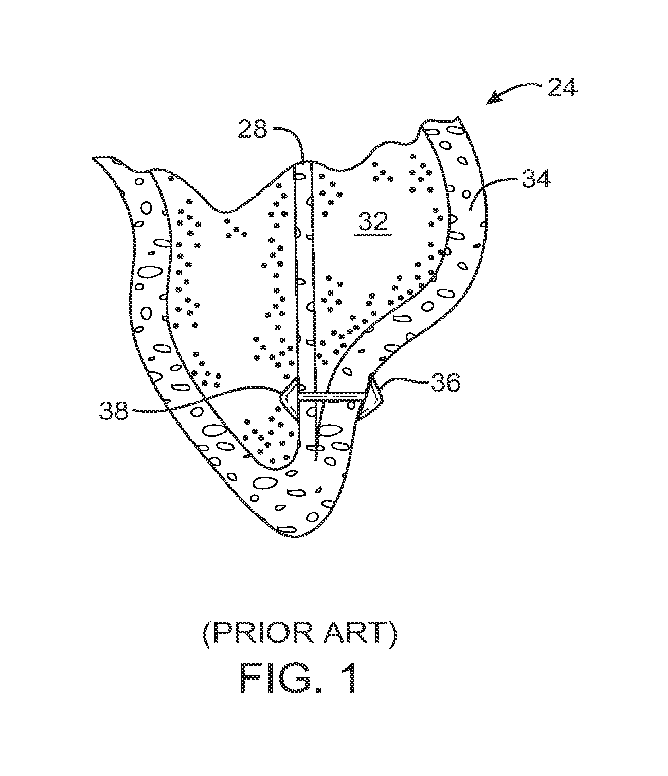
[0011]The present invention generally provides improved devices, systems, and methods for treating a heart of a patient. Embodiments of the invention may make use of structures which limit a size of a chamber of the heart, such as by deploying one or more tensile member to bring a wall of the heart and a septum of the heart toward each other (and often into contact). Therapeutic benefits of the implants may be enhanced by image-guided steering of the implant within the ventricle between penetration of the septum and wall. A plurality of tension members may help exclude scar tissue and provide a more effective remaining ventricle chamber. The implant may optionally be biodegradable, with the approximated surfaces of the septum and wall treated so as to induce the formation of adhesions. Antiproliferative agents or other drugs may be eluted from the implant to limit detrimental tissue responses and enhance the benefits of the implants for treatment of congestive heart failure and other disease states of the heart. Embodiments of this invention relate to devices and methods for completely off-pump treatment of congestive heart failure patients, particular sizing devices and methods for excluding infracted tissue and reducing ventricular volume. Some of the devices and methods described herein may be performed thoracoscopically off-pump and may be less traumatic to the patient than open chest and open heart surgical techniques.
[0014]The tension members will generally bring the wall and septum into engagement, and the separation between the tension members will allow the engagement to extend across at least a portion of the chamber. This engagement can effectively exclude regions of the wall and septum from the left ventricle. The anchors may extend laterally along the septum or wall towards each other (for example, having a width as measured extending toward an adjacent anchor that is greater than a height). The pattern of implants and anchors will generally be arranged to leave a remaining effective chamber that approximates the shape of a healthy heart chamber, avoids excessive thrombus-accumulating voids, and provides good effective pumping of blood therethrough.
[0015]In many embodiments, tissue near the first or second location may be engaged and characterized by a probe. If the characterized tissue does not appear suitable for formation of the penetration, the probe may be repositioned at a more suitable location. For example, a probe having a distal electrode surface may be advanced into contact with the tissue, and a pacing signal can be transmitted from the electrode. If the pacing signal is directly coupled to healthy, contractile heart tissue, the probe has effectively characterized the engaged tissue. As it may be desirable for the penetration to be formed in healthy tissue in some embodiments, engaged tissues which are not effectively paced by the applied signal may not be suitable for locating the anchor. In other embodiments, it may be desirable for the penetration to be formed in scar tissue which is not as susceptible to pacing, so that the implant may not fully exclude all scar tissue from the effective chamber. In either case, tissue characterization may help improve accuracy over deployment of the implant and efficacy of the therapy. The probe may comprise a perforation device, and may also be used to perforate the characterized tissue such as by energizing a bullet-shaped electrode surface of a steerable perforation device with electrosurgical energy.
[0016]The anchors will often be affixed by radially expanding the anchors and engaging axially-oriented surfaces of the anchors with tissue adjacent the perforations. For example, one or more of the anchors may comprise a plurality of arms defined by axial cuts in a tube. Radial expansion of the anchors may be effected by bending the arms radially outwardly, with the axially oriented surface comprising a first portion of each arm that extends perpendicular to the axis of the tube, and which is supported by a longer angled portion of the arm. In some embodiments, the axially-oriented surface may be supported by introducing a fluid into the anchor, with the fluid often being restrained by a bladder material similar to a balloon of a catheter balloon. Such a bladder may be used to support radially expanding arms as described above, or may be used as an anchor by itself The axially oriented surface does not necessarily have to be parallel to the axis of the tension member, and may angle radially outwardly while still providing sufficient axial tissue engagement for anchoring. The fill material may harden, reversibly or irreversibly, within the anchor. In some embodiments, the implant may release a bioactive material, such as by including a drug-eluting coating on at least a portion of the implant, by including pores in the bladder anchor which allow transmission of the bioactive agent from within the fill material, or the like. The agent may inhibit cell proliferation, enhance adhesion formation, and / or the like.
[0019]In another aspect, the invention provides a system for treating a heart. The heart has a first chamber bordered by a septum and a wall. The heart has a second chamber separated from the first chamber by the septum. The system comprises a plurality of implants. Each implant has an anchor, a wall anchor, and a tension member to apply tension between the septum and wall when the implant is deployed so as to bring the wall and septum into engagement. The implants together are configured to extend the engagement across a portion of the chamber (or all of the chamber) sufficiently to effectively exclude regions of both the wall and septum from the chamber.
[0021]In another aspect, the invention provides a system for treating a heart having a first chamber bordered by a septum and a wall, and a second chambered separated from the first chamber by the septum. The system comprises an implant having a septum anchor, a wall anchor, and a tension member to apply tension between the septum and wall when the implant is deployed so as to bring the wall and septum into engagement. At least one of the anchors has a small profile insertion configuration and large profile deployed configuration. The at least one anchor is radially expandable from the small profile configuration to the large profile configuration in situ so that an axially-oriented surface of the at least one anchor can anchor the implant to tissue of the heart.


