
Facilitating Temporal Comparison of Medical Images

a technology for temporal comparison and medical images, applied in image enhancement, image analysis, instruments, etc., can solve the problems of reducing the efficiency of radiologists, adversely affecting the experience of radiologists, so as to facilitate temporal comparison of breast mammograms

Inactive Publication Date: 2011-06-02
HOLOGIC INC
View PDF4 Cites 15 Cited by
  • Summary
  • Abstract
  • Description
  • Claims
  • Application Information

AI Technical Summary

Benefits of technology

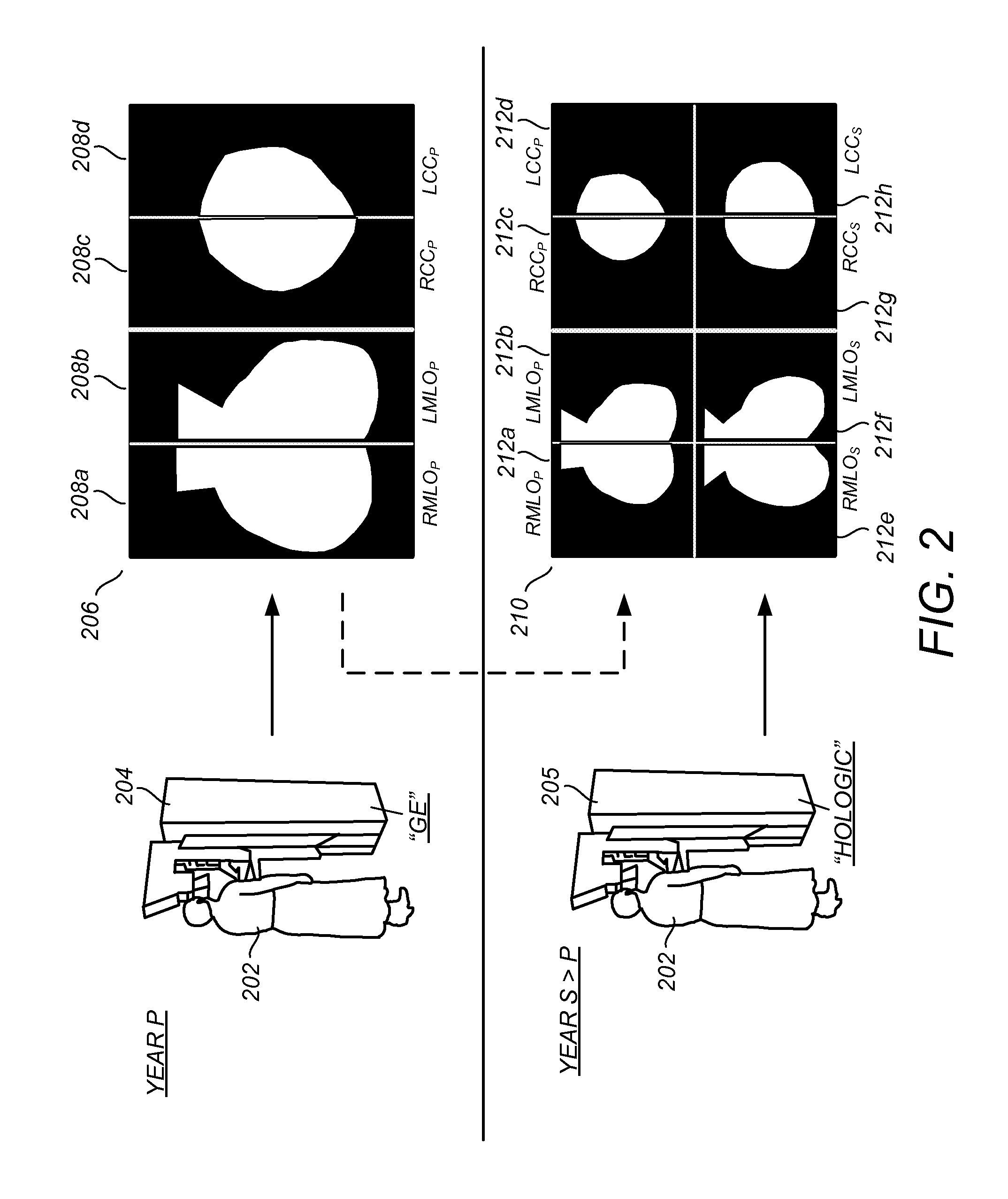
In one embodiment, a method for facilitating temporal comparison of breast mammograms by a viewer is provided. A prior mammographic image originating from a first mammogram acquisition system and having a first tissue distance per pixel is received, and a subsequent mammographic image of the breast originating from a second mammogram acquisition system is received. The second mammogram acquisition system is different than the first mammogram acquisition system and has a second tissue distance per pixel different than the first tissue distance per pixel. Without requiring a scale-adjusting viewer input, the prior and subsequent mammographic images are displayed for simultaneous viewing on a same mammogram display at an identical tissue distance per unit display distance.
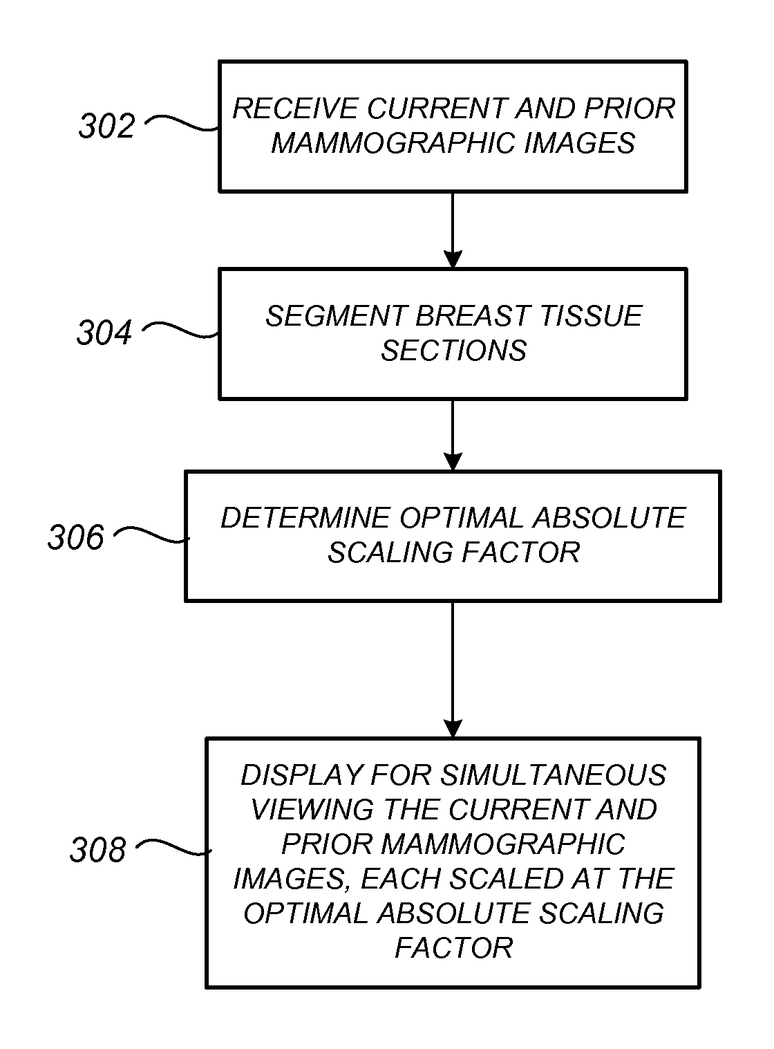
Also provided is a computer program product stored on a tangible medium for facilitating temporal comparison of breast mammograms by a viewer. The computer program product comprises computer code for receiving a first mammographic image of a breast originating from a first mammogram acquisition system and a temporally distinct second mammographic image of the breast originating from a second mammogram acquisition system different than the first mammogram acquisition system with respect to at least one of system manufacturer, detector type, detector size, and detector resolution. The computer program product further comprises computer code for segmenting each of the first and second mammographic images to identify a background section and a breast tissue section thereof. The computer program product still further comprises computer code for simultaneously displaying, without requiring a scale-adjusting or a window-adjusting viewer input, the first and second mammographic images at a same absolute spatial scale in commonly sized, substantially adjacent display windows on a mammogram display. Preferably, this “universal” absolute spatial scale for the entire temporal comparison display is a value for which (i) the breast tissue section of one of the mammographic images extends across a predetermined high percentage, such as 95%, of its display window in one of a heightwise and widthwise dimension, and extends across less than that predetermined high percentage in the other of the heightwise and widthwise dimensions, while also being the value for which the breast tissue section of the other mammographic image extends across less than the predetermined high percentage for both the heightwise and widthwise dimensions.
Also provided is a method for facilitating temporal comparison of digitally acquired breast mammograms by a viewer. The method comprises receiving, in a raw format, a prior mammographic image originating from a first digital mammogram acquisition system, and receiving a subsequent mammographic image of the breast originating from a second digital mammogram acquisition system different than the first digital mammogram acquisition system with respect to at least one of system manufacturer, detector characteristic, and display enhancement algorithm. If the first and second digital mammogram acquisition systems differ with respect to the detector characteristic, the pixel values of the raw-format prior mammographic image are remapped to estimate pixel values that would have resulted from acquiring the raw-format prior mammographic image from the second digital mammogram acquisition system. The method further comprises display-enhancing the raw-format prior mammographic image, which has been remapped if needed, according to one of an actual display enhancement algorithm and emulative display enhancement algorithm associated with the second digital mammogram acquisition system. Finally, the method further comprises displaying for simultaneous viewing the display enhanced remapped or non-remapped raw-format prior mammographic image and a display-enhanced version of the subsequent mammographic image processed according to the actual enhancement algorithm associated with the second digital acquisition system. Comparison between the prior and subsequent mammographic images is facilitated because, to an appealingly precise degree, both the prior and subsequent mammograms appear as if they were both acquired using the second mammogram acquisition system associated with the subsequent mammogram acquisition procedure.

Problems solved by technology

However, as discussed in the commonly-assigned U.S. Ser. 11 / 173,960, entitled “Displaying and Navigating Computer-Aided Detection Results on a Review Workstation,” which is incorporated by reference herein, problems can arise at the interface between (a) the amount of information available to a radiologist, and (b) the amount of information that can be usefully perceived by the radiologist in a reasonable amount of time.
Problems can arise in this side-by-side display that can adversely affect the radiologist experience, such as a need to repeatedly shift, re-window, or re-size the images, a need to repeatedly change brightness / contrast settings, etc., so that the prior and subsequent-year mammograms can be properly viewed for comparison.
This can bring about reduced radiologist efficiency, increased irritation or fatigue, or even missed detections or incorrect diagnoses resulting from the different size, scale, windowing, or otherwise different look of the side-by-side images not sufficiently corrected or correctable by the radiologist.

Method used


Image

  • Facilitating Temporal Comparison of Medical Images
  • Facilitating Temporal Comparison of Medical Images
  • Facilitating Temporal Comparison of Medical Images

Examples


Embodiment Construction

FIG. 1 illustrates a conceptual diagram of a medical imaging environment for which one or more of the preferred embodiments is particularly suited. Shown in FIG. 1 is a network 110, which may be a HIS / RIS (Hospital Information System / Radiology Information System) network, to which is coupled a film mammogram acquisition device 102, and a digital mammogram acquisition device 104. The film mammogram acquisition device 102 and digital mammogram acquisition device 104 are representative of a particular example of technology evolution that is / will be generally occurring in practical radiology environments, in which film-based mammogram systems are being phased out in favor of digital mammogram systems. Other situations to which the preferred embodiments described herein are particularly advantageous include, by way of nonlimiting example, (i) a scenario in which a patient visits a first mammography clinic in a first year that possesses a first mammogram acquisition system (film or digita...


PUM


Abstract

Methods, systems, and computer program products for facilitating temporal comparison of medical images is provided, with one exemplary application being for breast mammograms. In one embodiment, prior and subsequent mammographic images of a breast acquired using at least partially different mammogram acquisition systems are displayed for simultaneous viewing on a same mammogram display at an identical tissue distance per unit display distance without requiring a scale-adjusting viewer input. Also described are other embodiments for optimally scaling, windowing and / or otherwise advantageously processing and / or displaying prior and subsequent mammographic image sets in manners that facilitate temporal comparison therebetween.

Description

FIELDThis patent specification relates to medical imaging. More particularly, this patent specification relates to the processing and display of medical images of a body part acquired at different times using image acquisition equipment of different types and / or settings.BACKGROUNDProgress toward all-digital medical imaging environments has substantially increased the speed at which large amounts of medical image information can be accessed and displayed to a radiologist. As used herein, radiologist generically refers to a medical professional that analyzes medical images and makes clinical determinations therefrom, it being understood that such person might be titled differently, or might have differing qualifications, depending on the country or locality of their particular medical environment.One example of the increasing amount of information available to the radiologist relates to the temporal comparison of mammographic images, wherein years of prior mammographic images may be ...

Claims


Application Information

Patent Timeline
IPC IPC(8): G06T17/00G09G5/00
CPCG06T2207/30068G06T7/0014
Inventor ZINGARETTI, GABRIELEROEHRIG, JIMMY R.MARSHALL, JULIANTAO, ZHONG
Owner HOLOGIC INC
Learn More