
Bone fixation device, tools and methods

a bone fixation and bone technology, applied in the field of bone fixation devices, tools and methods for providing bone reconstruction and reinforcement, can solve the problems of pain or necrosis of bone, significant number of bone fractures, and take several months for bone to heal

Inactive Publication Date: 2011-04-14
ARTHREX
View PDF93 Cites 97 Cited by
  • Summary
  • Abstract
  • Description
  • Claims
  • Application Information

AI Technical Summary

Benefits of technology

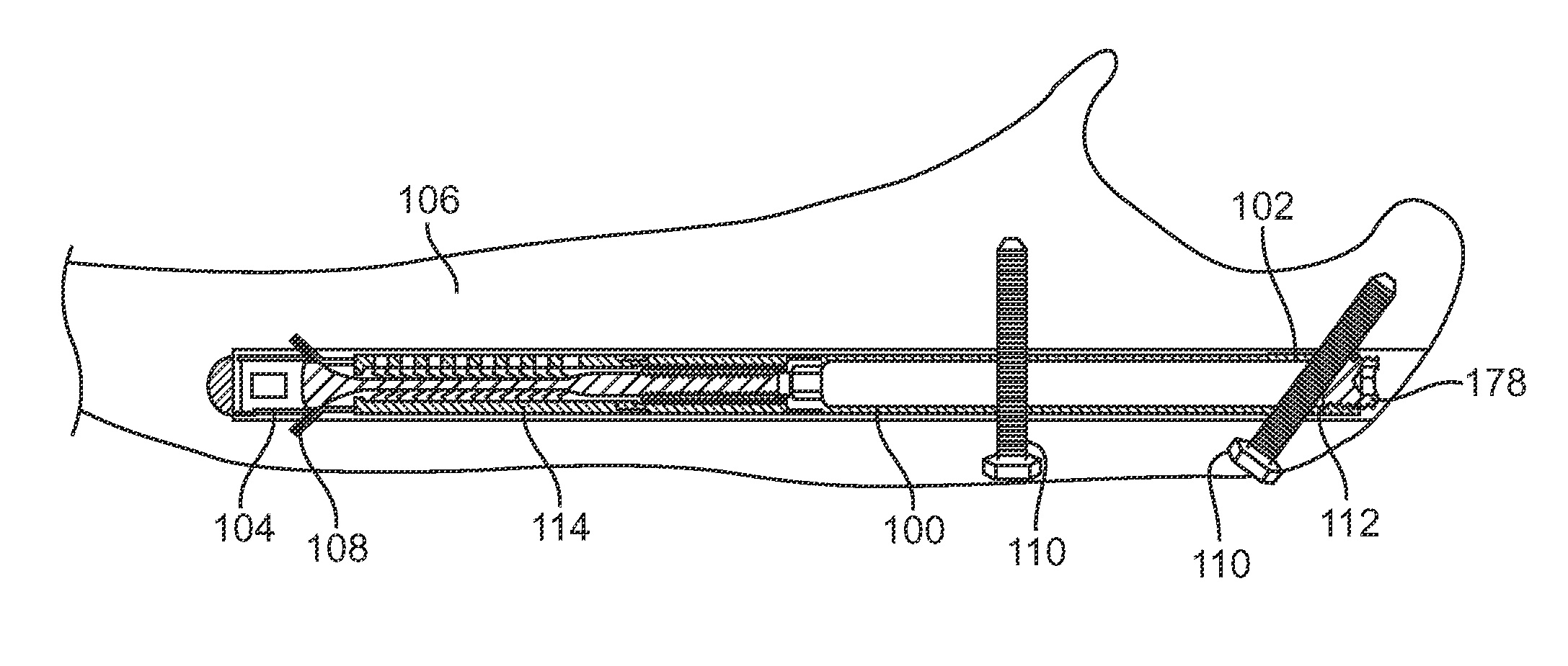
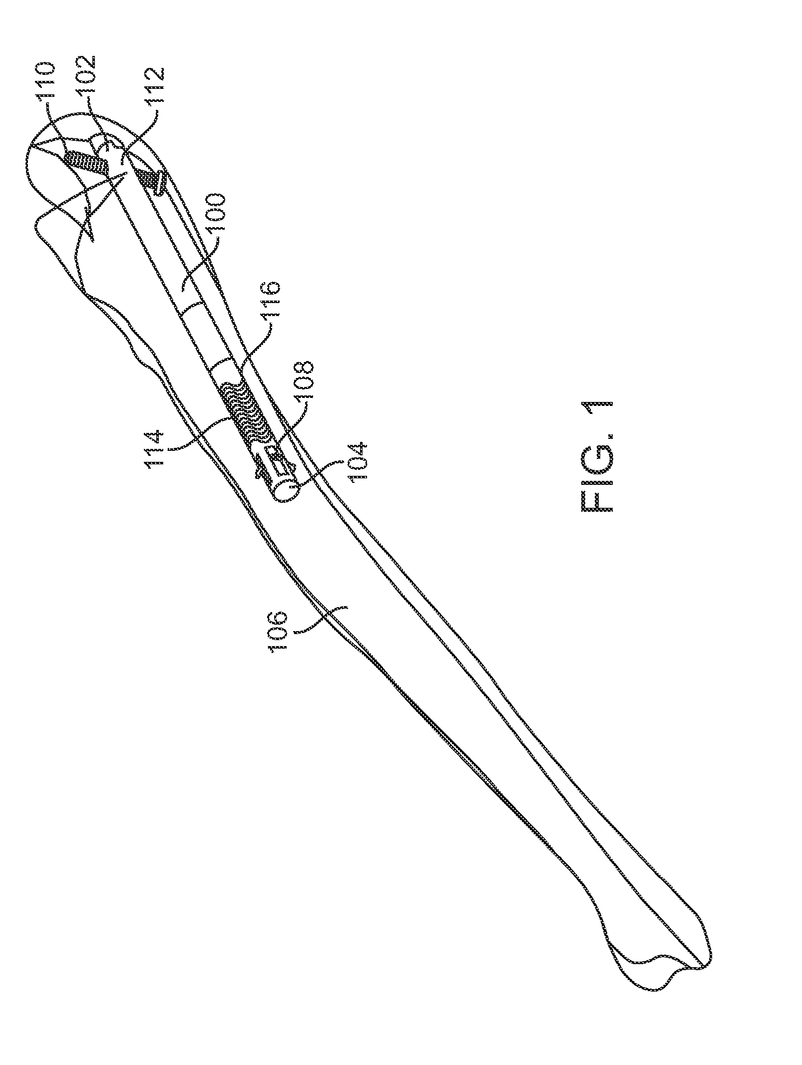
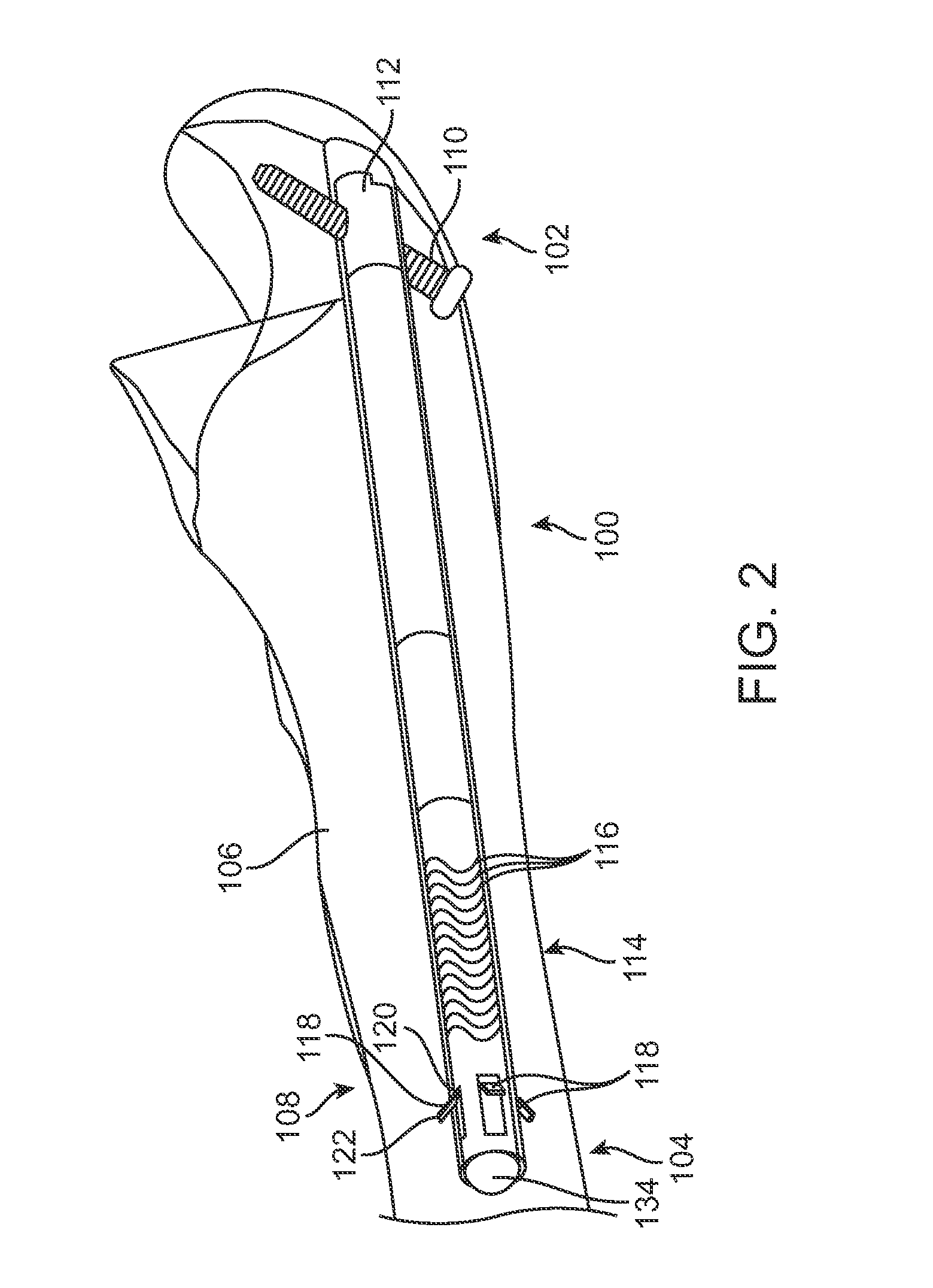
[0014]One embodiment of the present invention provides a low weight to volume mechanical support for fixation, reinforcement and reconstruction of bone or other regions of the musculo-skeletal system in both humans and animals. The method of delivery of the device is another aspect of the invention. The method of delivery of the device in accordance with the various embodiments of the invention reduces the trauma created during surgery, decreasing the risks associated with infection and thereby decreasing the recuperation time of the patient. The framework may in one embodiment include an expandable and contractible structure to penult re-placement and removal of the reinforcement structure or framework.
[0025]Embodiments of the invention also provide deployment tools with a tool guide for precise alignment of one or more bone screws with the fracture fixation device. These embodiments also provide bone screw orientation flexibility so that the clinician can select an orientation for the bone screw(s) that will engage the fixation device as well as any desired bone fragments or other bone or tissue locations.

Problems solved by technology

However, with an increasingly aging population, osteoporosis has become more of a significant medical concern in part due to the risk of osteoporotic fractures.
Also, sports and work-related accidents account for a significant number of bone fractures seen in emergency rooms among all age groups.
This can take several months for the bone to heal and for the patient to remove the cast before resuming normal activities.
Further, micro-motion between the bone and the IM nail may cause pain or necrosis of the bone.
This requires a subsequent surgery with all of the complications and risks of a later intrusive procedure.
In general, rigid IM rods or nails are difficult to insert, can damage the bone and require additional incisions for cross-screws to attach the rods or nails to the bone.
Such IM nails require inflating the rod with very high pressures, endangering the surrounding bone.
Furthermore, the external fixation is cosmetically intrusive, bulky, and prone to painful inadvertent manipulation by environmental conditions such as, for example, bumping into objects and laying on the device.

Method used


Image

  • Bone fixation device, tools and methods
  • Bone fixation device, tools and methods
  • Bone fixation device, tools and methods

Examples


Embodiment Construction

[0060]By way of background and to provide context for the invention, it may be useful to understand that bone is often described as a specialized connective tissue that serves three major functions anatomically. First, bone provides a mechanical function by providing structure and muscular attachment for movement. Second, bone provides a metabolic function by providing a reserve for calcium and phosphate. Finally, bone provides a protective function by enclosing bone marrow and vital organs. Bones can be categorized as long bones (e.g. radius, femur, tibia and humerus) and flat bones (e.g. skull, scapula and mandible). Each bone type has a different embryological template. Further each bone type contains cortical and trabecular bone in varying proportions. The devices of this invention can be adapted for use in any of the bones of the body as will be appreciated by those skilled in the art.

[0061]Cortical bone (compact) forms the shaft, or diaphysis, of long bones and the outer shell...


PUM


Abstract

A bone fixation device is provided with an elongate body having a longitudinal axis and having a first state in which at least a portion of the body is flexible and a second state in which the body is generally rigid, an actuateable gripper disposed at one or more locations on the elongated body, a hub located on a proximal end of the elongated body, and an actuator operably connected to the gripper(s) to deploy the gripper(s) from a retracted configuration to an expanded configuration. Methods of repairing a fracture of a bone are also disclosed. One such method comprises inserting a bone fixation device into an intramedullary space of the bone to place at least a portion of an elongate body of the fixation device in a flexible state on one side of the fracture and at least a portion of a hub on another side of the fracture, and operating an actuator to deploy at least one gripper of the fixation device to engage an inner surface of the intramedullary space to anchor the fixation device to the bone. Various hub designs are disclosed that may be used in combination with other fixation device components.

Description

CROSS-REFERENCE TO RELATED APPLICATIONS[0001]This application claims benefit to U.S. Provisional Application No. 61 / 138,920 filed Dec. 18, 2008 and entitled, “BONE FIXATION DEVICE, TOOLS AND METHODS.”INCORPORATION BY REFERENCE[0002]This application is related to: U.S. application Ser. No. 11 / 383,269 filed May 15, 2006, entitled, “MINIMALLY INVASIVE ACTUABLE BONE FIXATION DEVICES”; U.S. application Ser. No. 11 / 944,366 filed Nov. 21, 2007, entitled, “FRACTURE FIXATION DEVICE, TOOLS AND METHODS”; U.S. application Ser. No. 12 / 482,388, filed Jun. 10, 2009, entitled, “FRACTURE FIXATION DEVICE, TOOLS AND METHODS”; U.S. application Ser. No. 12 / 482,395 filed on Jun. 10, 2009 entitled, “FRACTURE FIXATION DEVICE, TOOLS AND METHODS” and U.S. application Ser. No. 12 / 482,406 filed on Jun. 10, 2009, entitled, “FRACTURE FIXATION DEVICE, TOOLS AND METHODS,” all of which are incorporated herein by reference. All publications and patent applications mentioned in this specification are herein incorpora...

Claims


Application Information

Patent Timeline
Patent Type & Authority Applications(United States)
IPC IPC(8): A61B17/56
CPCA61B17/1725A61B17/68A61B17/7208A61B17/72A61B17/7266A61B17/7291A61B17/86A61B17/7225
Inventor MAZUR, KAL U.MCDANIEL, STEPHEN R.PHAM, TRUNG HONELSON, CHARLES L.GUNTHER, STEPHEN B.SARAVIA, HEBERCOLEMAN, ROBERT G.
Owner ARTHREX
Learn More