Method and system for monitoring skeletal defects
a technology for skeletal defects and skeletal bones, applied in the field of skeletal defects monitoring, can solve the problems of large equipment, specialized facilities, high cost, and current methods of detection including x-rays, mris and cts for detecting bone fractures, and achieve rapid and inexpensive detection and diagnosis, improve detection accuracy, and improve the effect of bone fracture detection
- Summary
- Abstract
- Description
- Claims
- Application Information
AI Technical Summary
Benefits of technology
Problems solved by technology
Method used
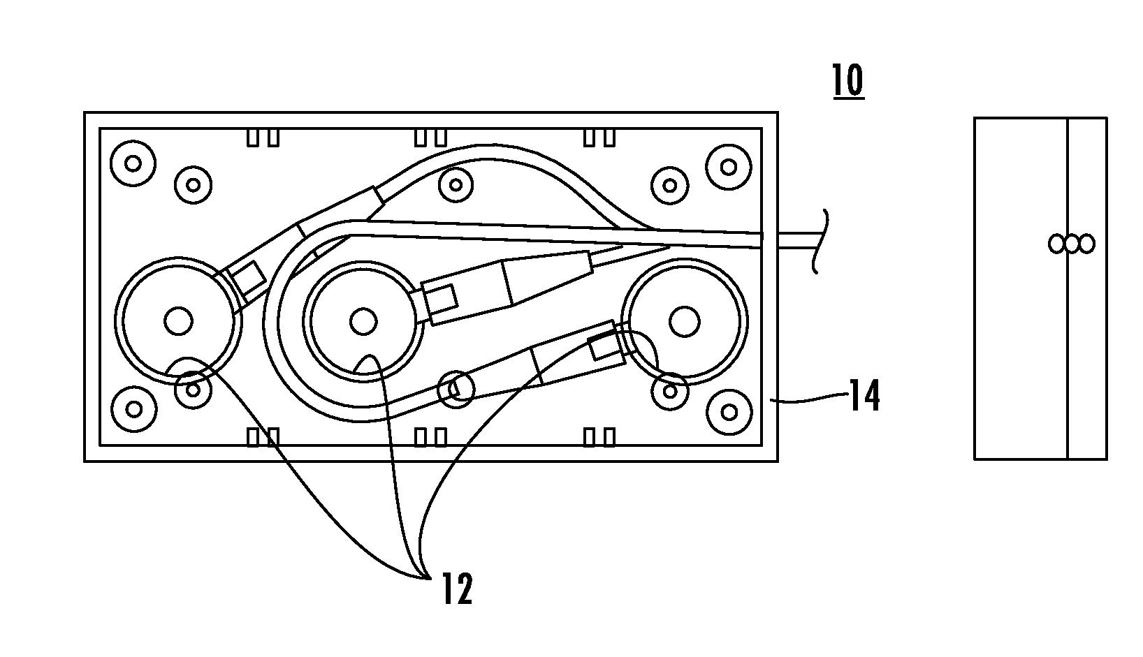
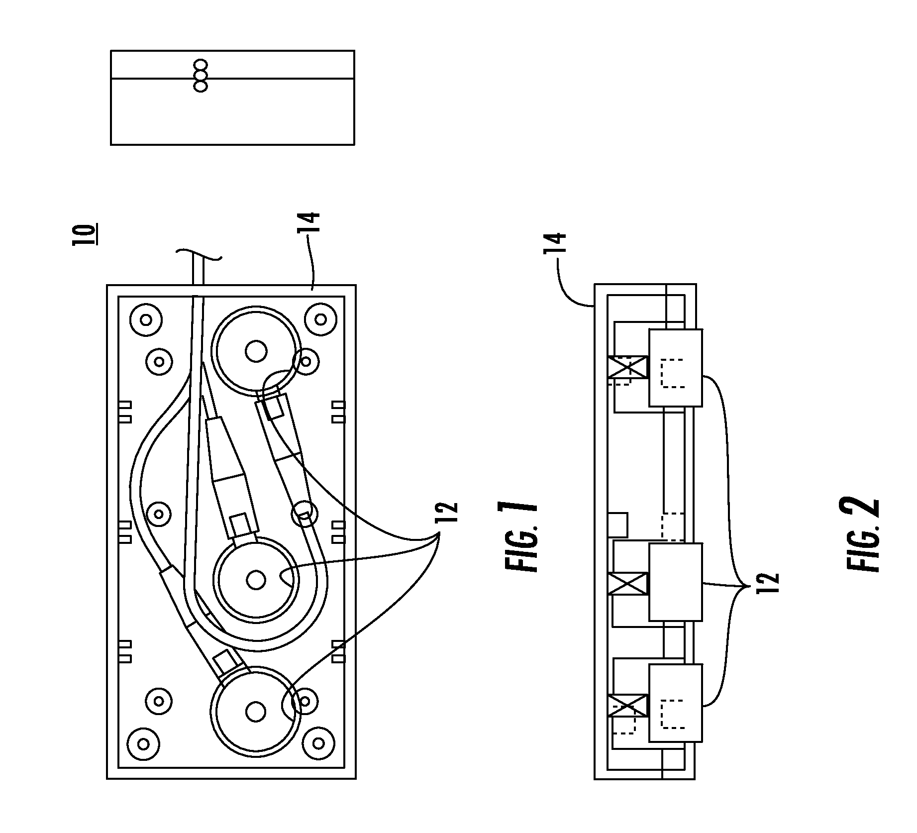
Image

Examples
example 1
Bovine Femur Testing
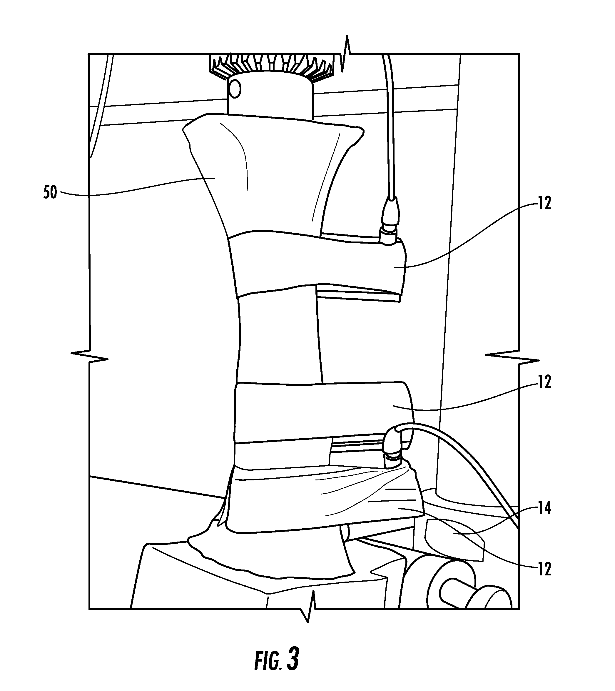
[0029]Tests were performed on bone with precise cuts using a fine saw to mimic fatigue fractures in the bones cortical surface (hard outer layer) of varying depths and lengths using device 10 upon application to soft tissues as shown in FIG. 3. Five parameters of axial transmission were evaluated to establish the transducer and signal combination which yields the optimal detection and monitoring of fatigue fractures.
example 2
Evaluation and Correlation
[0030]Experimental data was gathered for the evaluation of the five parameters of axial transmission, shown in FIG. 4, by using cross correlation techniques which established the detection and monitoring of fatigue fractures. These tests provide comprehensive results for simulated fatigue fractures of various sizes. The results define the combination of transducer frequency and acoustic parameters that have the greatest sensitivity and specificity to the presence of a fracture. The highest sensitivity is defined as the largest change in a parameter's value for the smallest change in fracture size. The highest specificity is defined as the accurate identification and characterization of a fracture. This data provides the critical technical parameters for defining the ultrasonic transducers' characteristics and configuration.
example 3
Test Results
[0031]FIG. 9A-9E provides graphs made while performing tests on the bovine femur, as shown in FIG. 3, using system 100, as shown in FIG. 4. Cuts were made by surgically increasing the depth of cut in the cortical portion of the bone and charting the results of the five parameters.
[0032]Each graph shows, reading left to right horizontally, the deepest cut on the left of the chart, to the smallest cut and uncut bone on the right of the chart in millimeter depth (5 to 10 mm). As shown, the signal decreases as the depth of the cut increases, on the left of the chart, giving a well defined change as the signal increases with reduction in cut depth at the right of the chart in FIGS. 9A-9E. However, in the case of Flight Time, graph is reversed in FIG. 9C.
[0033]These results have shown that system 100 provides improved definition in signal information and thus can indicate a small cut which mimics a stress fracture in all the five parameters measured.
[0034]FIG. 9A illustrates g...
PUM
 Abstract
Description
Claims
Application Information
 - R&D
- Intellectual Property
- Life Sciences
- Materials
- Tech Scout
- Unparalleled Data Quality
- Higher Quality Content
- 60% Fewer Hallucinations
Browse by: Latest US Patents, China's latest patents, Technical Efficacy Thesaurus, Application Domain, Technology Topic, Popular Technical Reports.
© 2025 PatSnap. All rights reserved.Legal|Privacy policy|Modern Slavery Act Transparency Statement|Sitemap|About US| Contact US: help@patsnap.com