


Quick Research
Generate reliable direction feasibility study reports for your R&D in just a few steps.

Technical Q&A
Discover and master advanced knowledge NOW. Basics, ideas, possibilities, all at once.

Find Solutions
As an expert in R&D theories, this can generate solutions to your technical problems instantly.

Evaluate Feasibility
Analyze your overall solution with one click, know your potential R&D risks in advance.

Monitor Landscape
Get weekly tech updates, stay abreast of the latest tech innovations and key insights.
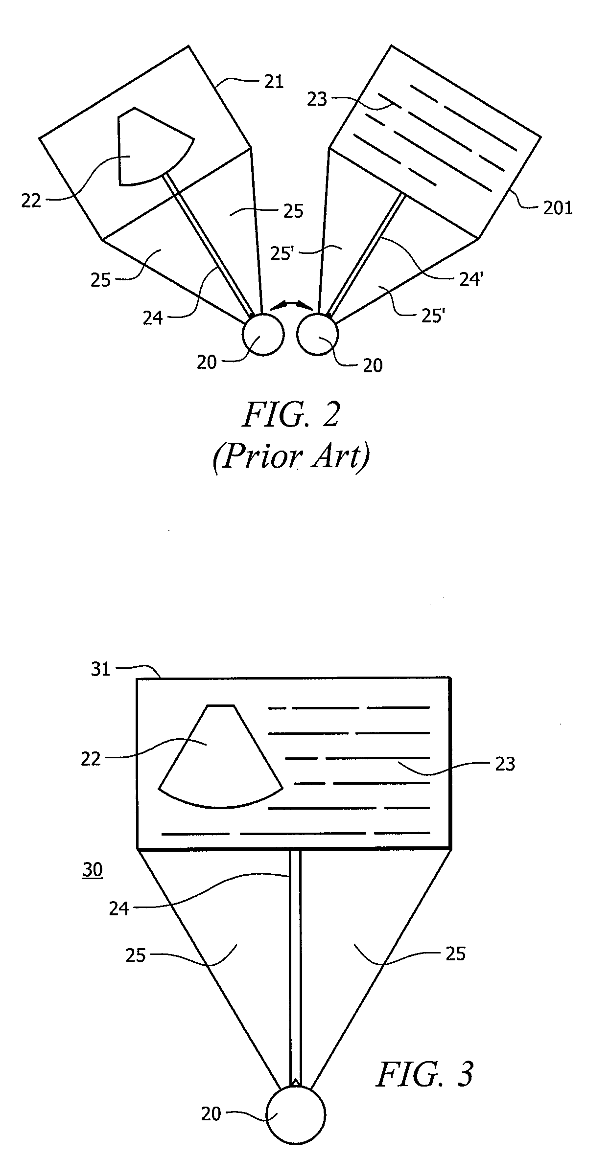
Systems and Methods for the Display of Ultrasound Images
a technology for ultrasound images and patient records, applied in ultrasonic/sonic/infrasonic diagnostics, instruments, applications, etc., can solve the problems of affecting the efficiency and accuracy of sonographers, the sonographer is subject to making mistakes using this method, and the efficiency of sonographers is affected, so as to facilitate less reliance on memory and improve the efficiency of sonographers
- Summary
- Abstract
- Description
- Claims
- Application Information
AI Technical Summary
Benefits of technology
Problems solved by technology
Method used
Image


Examples
Embodiment Construction
[0014]FIG. 1 shows work flow 10 of a sonographer doing an ultrasound examination of a patient. In process 101, the sonographer prepares to conduct the examination. This may include entering patient identification and the procedure being performed into the database of an ultrasound device. Preparation may also involve applying a gel to the area of the body to be scanned. In process 102, the sonographer uses the transducer—the component of the ultrasound machine that emits the ultrasound—to prescan a part of the patient's body by moving the activated transducer against the body. In process 103, the sonographer may then apply more gel to the relevant part of the body and performs a complete scan of the relevant part of the body.
[0015]As the sonographer views the ultrasound images in process 103, the sonographer may determine that the ultrasound image on the screen shows something of interest. In process 104, the sonographer freezes the ultrasound image and measures portions of the ultr...
PUM
 Abstract
Description
Claims
Application Information
 - R&D Engineer
- R&D Manager
- IP Professional
- Industry Leading Data Capabilities
- Powerful AI technology
- Patent DNA Extraction
Browse by: Latest US Patents, China's latest patents, Technical Efficacy Thesaurus, Application Domain, Technology Topic, Popular Technical Reports.
© 2024 PatSnap. All rights reserved.Legal|Privacy policy|Modern Slavery Act Transparency Statement|Sitemap|About US| Contact US: help@patsnap.com