
Multimodal Spectroscopic Systems and Methods for Classifying Biological Tissue

a multi-modal spectroscopic and biological tissue technology, applied in the field of medical imaging systems, can solve the problems of small tissue diagnosis sites, relative non-expensive, etc., and achieve the effect of small sites and non-expensiv

Active Publication Date: 2009-12-24
RGT UNIV OF MICHIGAN
View PDF2 Cites 24 Cited by
  • Summary
  • Abstract
  • Description
  • Claims
  • Application Information

AI Technical Summary

Benefits of technology

[0013]The systems and methods described herein are well-suited to be used in conjunction with or as a substitute for random biopsies, since the optical systems and methods are non-invasive, do not require tissue removal, and can be performed in-vivo. Moreover, they are fast (can be applied in real time), are relatively non-expensive, are able to work on microscopic scale, and thus can find very small sites for tissue diagnosis, which may be missed by random biopsies. The systems and methods herein are also well-suited to be used in endoscopic ultrasound-guided procedures and apparatus, or delivered through an endoscope or needle by a trained professional.

Problems solved by technology

Moreover, they are fast (can be applied in real time), are relatively non-expensive, are able to work on microscopic scale, and thus can find very small sites for tissue diagnosis, which may be missed by random biopsies.

Method used


Image

  • Multimodal Spectroscopic Systems and Methods for Classifying Biological Tissue
  • Multimodal Spectroscopic Systems and Methods for Classifying Biological Tissue
  • Multimodal Spectroscopic Systems and Methods for Classifying Biological Tissue

Examples


Embodiment Construction

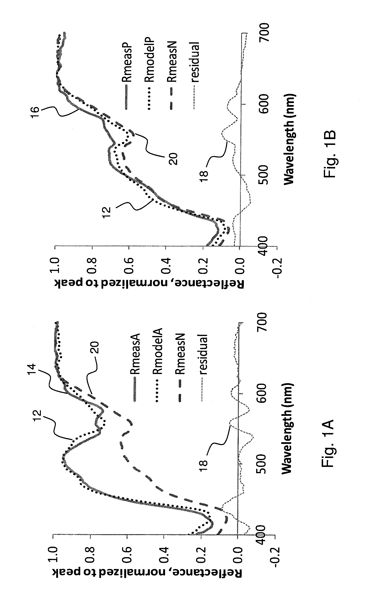
[0035]Some embodiments of the invention employ multimodal optical spectroscopic systems and methods to obtain data from biological tissue and compare the data with preset criteria configured to aid in the diagnosis of the tissue health or condition, wherein the preset criteria relates the data with the most probable attributes of the tissue. The multimodal spectroscopic systems employed may include fluorescence spectroscopy, reflectance spectroscopy and time-resolved spectroscopy, among others.

[0036]In some embodiments, data obtained through multimodal optical spectroscopy is correlated with the results of a microscopic histological examination of a normal tissue sample to develop the preset criteria by which further tissue samples are to be assessed. In particular, the preset criteria may be based on a relationship between spectral data and the histological aspects of the tissue which are most likely to be indicative of a specific attribute so as to lead to a unique classification ...


PUM


Abstract

Multimodal optical spectroscopy systems and methods produce a spectroscopic event to obtain spectroscopic response data from biological tissue and compare the response data with preset criteria configured to correlate the measured response data and the most probable attributes of the tissue, thus facilitating classification of the tissue based on those attributes for subsequent biopsy or remedial measures as necessary.

Description

CROSS-REFERENCE TO RELATED APPLICATION[0001]The instant application claims priority to U.S. Provisional Patent Application Ser. No. 61 / 058,966, filed Jun. 5, 2008, the entire specification of which is expressly incorporated herein by reference.STATEMENT REGARDING FEDERALLY SPONSORED RESEARCH OR DEVELOPMENT[0002]The invention was made with U.S. Government support, in whole or in part, from NIH grant no. CA-114542 and NIH / NCI grant no. ______. The U.S. Government has certain rights in the invention.FIELD OF THE INVENTION[0003]The present invention generally relates to medical imaging systems and in particular, to employing multimodal spectroscopy in the diagnosis of biological tissue.BACKGROUND OF THE INVENTION[0004]The following description of the background of the invention is provided to aid in understanding the invention, but is not admitted to describe or constitute prior art to the invention. The contents of the articles, patents, and patent applications, and all other documents...

Claims


Application Information

Patent Timeline
IPC IPC(8): C12Q1/02C12M1/34
CPCA61B5/0071G01N2201/1293A61B5/0084A61B5/415A61B5/418A61B5/7264A61B5/7267G01J3/10G01J3/4406G01N21/474G01N21/6408G01N21/645G01N21/6486G01N33/57438G01N2021/4742G01N2021/6484A61B5/0075G16H50/20
Inventor MYCEK, MARY-ANNCHANDRA, MALAVIKASCHEIMAN, JAMESWILSON, ROBERT H.SIMEONE, DIANEMCKENNA, BARBARAPURDY, JULIANNE
Owner RGT UNIV OF MICHIGAN
Learn More