Methods for Diagnosing Cancer Using Samples Collected From A Central Vein Location or an Arterial Location

a central vein or arterial location technology, applied in biochemistry equipment and processes, laboratory glassware, instruments, etc., can solve the problems of many cancers going undetected and damaging healthy tissu

Inactive Publication Date: 2009-09-17
CELLECTIVE DX CORP
View PDF6 Cites 25 Cited by
  • Summary
  • Abstract
  • Description
  • Claims
  • Application Information

AI Technical Summary

Benefits of technology

[0006]The invention provides for methods to selectively concentrate one or more rare particles from an organism comprising: applying a blood sample taken from a jugular vein, a femoral vein, a subclavian vein, an artery or a heart to a device that selectively enriches the one or more rare particles. The artery can be a radial artery, an ulnar artery, a brachial artery, a femoral artery, a carotid artery, a subclavian artery or a brachiocephalic artery. The method can be used for blood samples taken from human or non-human animals. The rare particles can be a variety of rare particles, including circulating tumor cells, circulating stem cells or fragments thereof.

Problems solved by technology

Metastasis results in the formation of secondary tumors at multiple sites, damaging healthy tissue.
Despite decades of advances in cancer diagnosis and therapy, many cancers continue to go undetected until late in their development.

Method used


Image

  • Methods for Diagnosing Cancer Using Samples Collected From A Central Vein Location or an Arterial Location
  • Methods for Diagnosing Cancer Using Samples Collected From A Central Vein Location or an Arterial Location
  • Methods for Diagnosing Cancer Using Samples Collected From A Central Vein Location or an Arterial Location

Examples


example 1

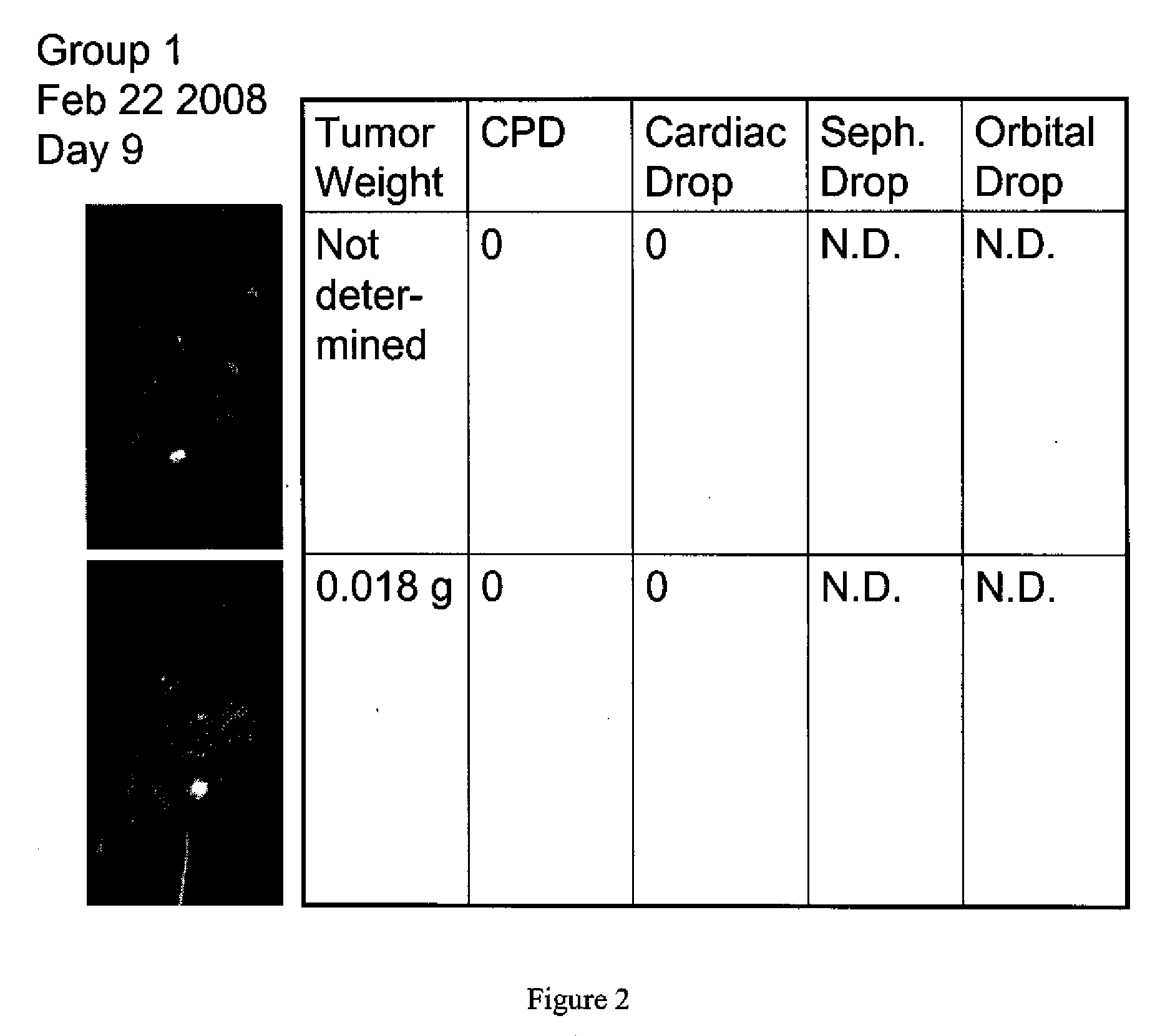
[0033]A mouse study evaluating three different bleed sites were for recovery of circulating tumor cells (CTC) was performed.

[0034]Sample Collection

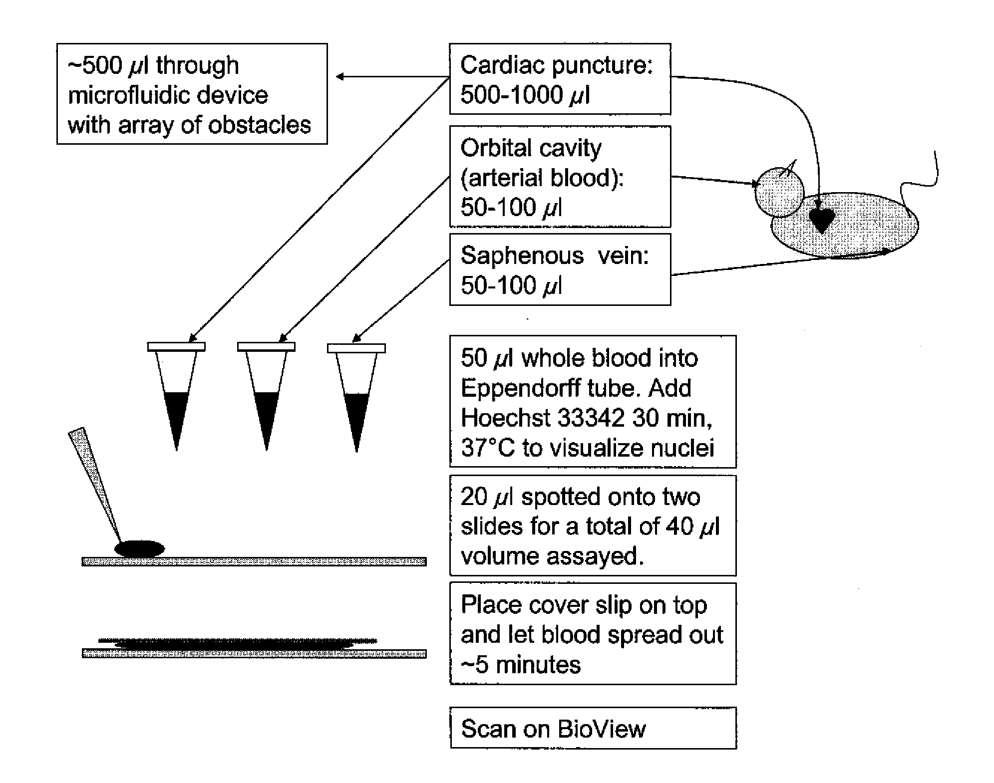
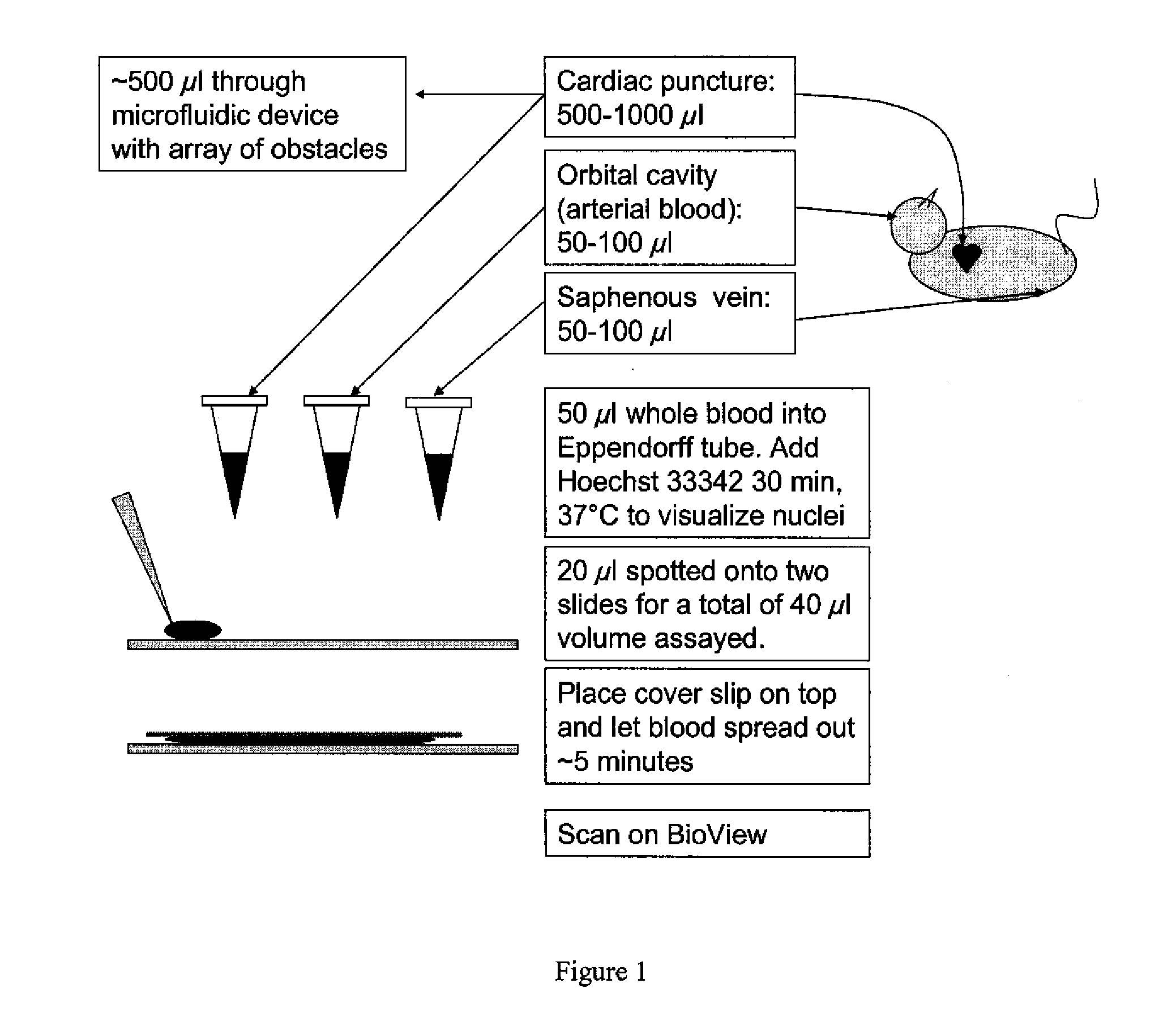
[0035]At two time points, three groups of mice, including two positive control mice and one negative control mice, were sampled. Positive control mice were subjected to injection of GFP producing tumors. Blood samples were obtained via a saphenous vein, a retro-orbital bleed (arterial blood) and cardiac puncture. An illustration depicting the locations for blood sampling is shown in FIG. 1. One group of mice was terminated and primary tumor and metastatic tissue was collected.

[0036]Analysis

[0037]Each blood sample was evaluated for amount of circulating tumor cells prior to enrichment using a photomicroscope. For the cardiac puncture samples, circulating tumor cells were selectively enriched from the blood sample. The enrichment was performed using a microfluidic device with a two-dimensional array of obstacles that contained pinch points ...


PUM

PropertyMeasurementUnit
weightaaaaaaaaaa
affinityaaaaaaaaaa
sizeaaaaaaaaaa

Abstract

The invention encompasses methods for selectively enriching in rare particles from blood samples harvested from the vein jugular vein the femoral vein, the subclavian vein, or an artery. These rare particles can be circulating tumor cells, circulating stem cells, or fragments thereof. Blood samples harvested from different sources can contain higher or lower concentrations of rare particles. The rare particles can be enriched by applying the blood samples to a microfluidic device with a two dimensional array of obstacles.

Description

TECHNICAL FIELD[0001]The invention is related to medical diagnostics and methods of sampling blood for enrichment of rare particles.BACKGROUND[0002]Cancer is a disease marked by the uncontrolled proliferation of abnormal cells. In normal tissue, cells divide and organize within the tissue in response to signals from surrounding cells. Cancer cells do not respond in the same way to these signals, causing them to proliferate and, in many organs, form a tumor. As the growth of a tumor continues, genetic alterations may accumulate, manifesting as a more aggressive growth phenotype of the cancer cells. If left untreated, metastasis, the spread of cancer cells to distant areas of the body by way of the lymph system or bloodstream, may ensue. Metastasis results in the formation of secondary tumors at multiple sites, damaging healthy tissue. Most cancer death is caused by such secondary tumors.[0003]Despite decades of advances in cancer diagnosis and therapy, many cancers continue to go und...

Claims


Application Information

Patent Timeline
Patent Type & Authority Applications(United States)
IPC IPC(8): C12Q1/02C12N5/06
CPCB01L3/502761B01L2300/0681B01L2300/0816G01N33/57488B01L2300/16B01L2400/086B01L2300/0877
Inventor KOPF-SILL, ANNE R.
Owner CELLECTIVE DX CORP
Why Eureka
Try Eureka