
Intraluminal tissue markers

a tissue marker and intraluminal technology, applied in the field of intraluminal tissue markers, can solve the problems of intrinsic danger of possibly injecting dye into the peritoneal cavity, tissue cannot be removed, intended site may become obscure,

Inactive Publication Date: 2009-09-03
ETHICON ENDO SURGERY INC
View PDF29 Cites 152 Cited by
  • Summary
  • Abstract
  • Description
  • Claims
  • Application Information

AI Technical Summary

Benefits of technology

[0061]One skilled in the art will appreciate further features and advantages of the invention based on the above-described embodiments. Accordingly, the invention is not to be limited by what has been particularly shown and described, except as indicated by the appended claims. All publications and references cited herein are expressly incorporated herein by reference in their entirety.

Problems solved by technology

However, in some cases the tissue cannot be removed during the colonoscopy, and thus must be removed in a subsequent surgical procedure.
However, such a procedure includes the intrinsic danger of possibly injecting dye into the peritoneal cavity.
In addition, the injected marker may also spread so widely that the intended site may become obscured.

Method used


Image

  • Intraluminal tissue markers
  • Intraluminal tissue markers
  • Intraluminal tissue markers

Examples


Embodiment Construction

[0026]Certain exemplary embodiments will now be described to provide an overall understanding of the principles of the structure, function, manufacture, and use of the devices and methods disclosed herein. One or more examples of these embodiments are illustrated in the accompanying drawings. Those skilled in the art will understand that the devices and methods specifically described herein and illustrated in the accompanying drawings are non-limiting exemplary embodiments and that the scope of the present invention is defined solely by the claims. The features illustrated or described in connection with one exemplary embodiment may be combined with the features of other embodiments. Such modifications and variations are intended to be included within the scope of the present invention.

[0027]The present invention generally provides methods and devices for marking a target tissue to be subsequently located for removal from a body, for diagnosis, for treatment, or for other purposes. ...


PUM


Abstract

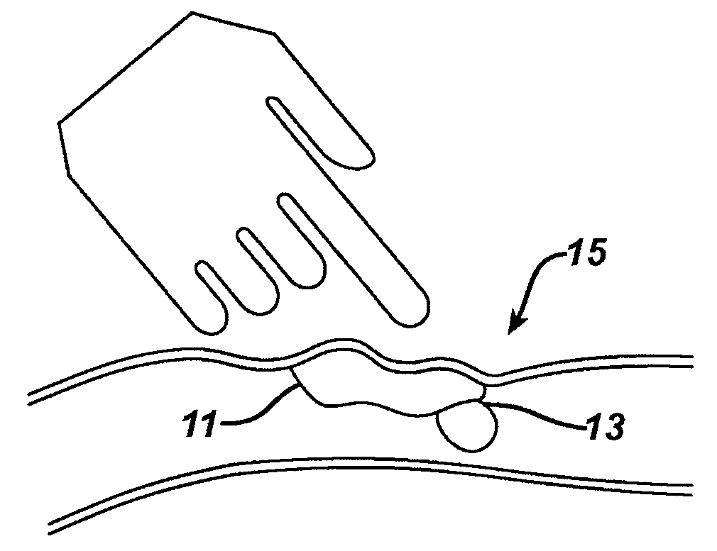
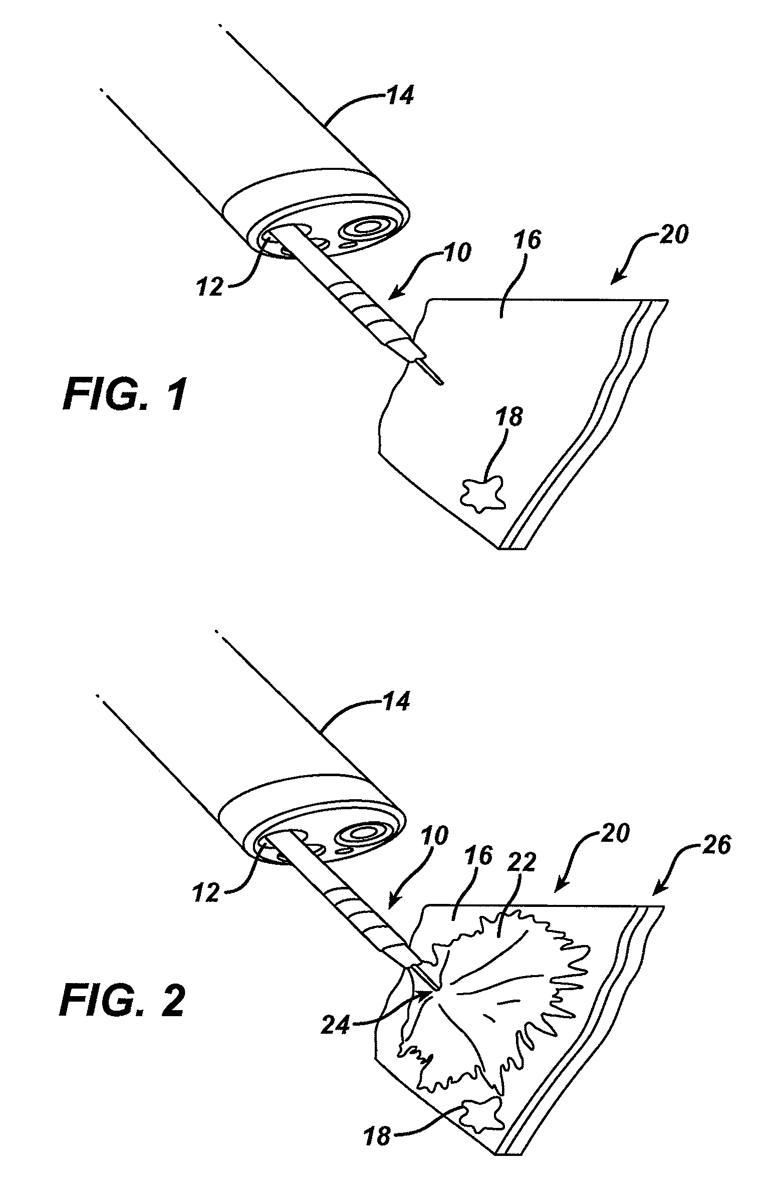
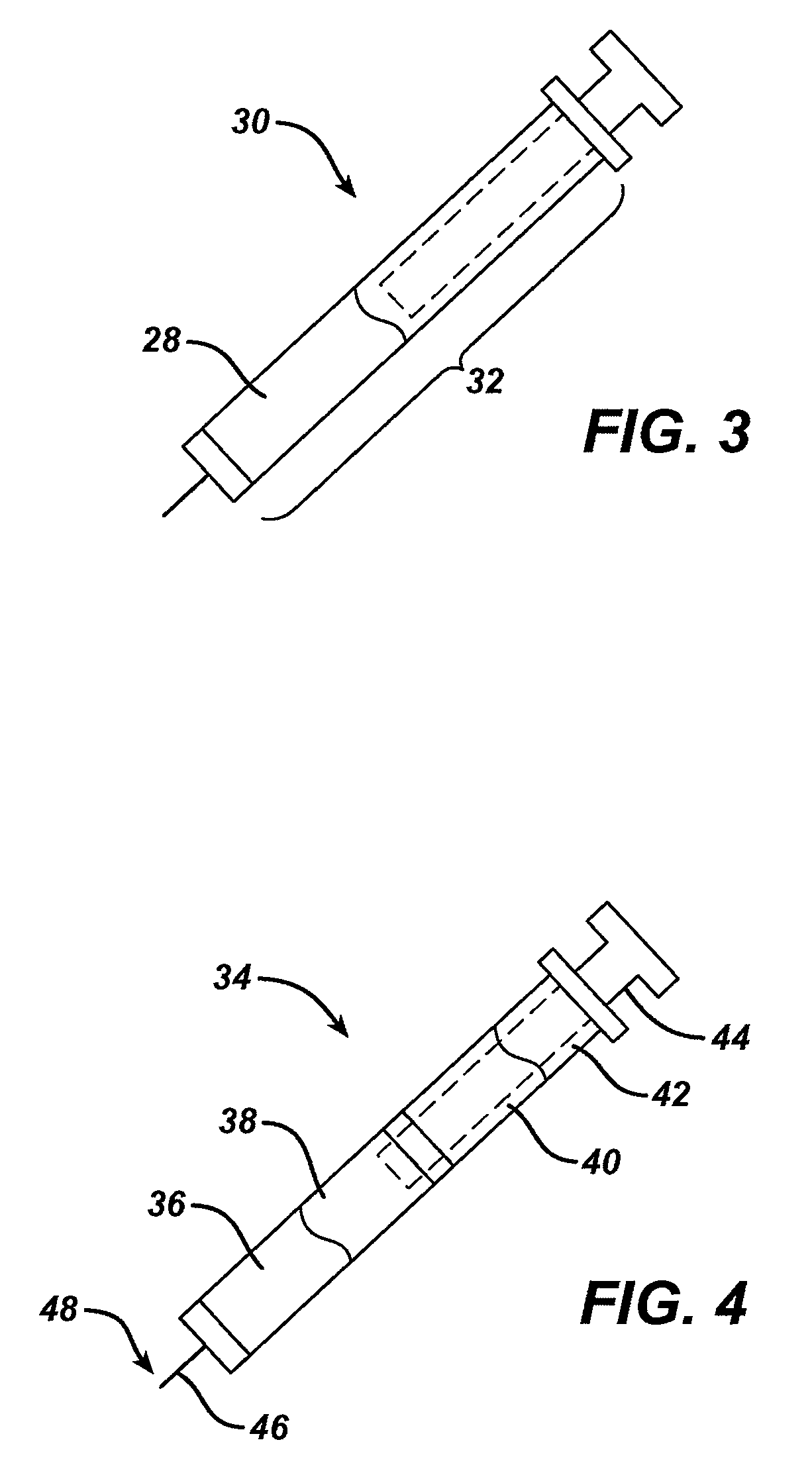
Methods and devices are provided for marking tissue to be subsequently located for removal from a body or for other examination. In general, a marker is provided that can be delivered to a target tissue. In one embodiment, the marker can include a solution having a visual marking component and a palpably identifiable tactile marking component. The marker can remain in the body and be subsequently visually and / or palpably identified to locate the target tissue.

Description

FIELD OF THE INVENTION[0001]The present invention relates to intraluminal tissue markers and methods for marking tissue intraluminally.BACKGROUND OF THE INVENTION[0002]Colonoscopy is an outpatient procedure in which the rectum and the inside of the lower large intestine (colon) are examined. Colonoscopies are commonly used to evaluate bowel disorders, rectal bleeding or polyps (usually benign growths) found on contrast x-rays. Colonoscopies are also performed to screen people over age 50 for colon and rectal cancer. During a colonoscopy, a physician uses a colonoscope (a long, flexible instrument about ½ inch in diameter) to view the lining of the colon. The colonoscope is inserted through the rectum and advanced to the large intestine.[0003]If necessary during a colonoscopy, small amounts of tissue can be removed for analysis (called a biopsy) and polyps can be identified and removed. In many cases, colonoscopy allows accurate diagnosis and treatment without the need for a major op...

Claims


Application Information

Patent Timeline
IPC IPC(8): A61B19/00
CPCA61B5/0071A61B5/0084A61B19/54A61B2019/5408A61B2019/5433A61B2019/5495A61B2019/5441A61B2019/545A61B2019/5462A61B2019/547A61B2019/5487A61B2019/5437A61B90/39A61B2090/3908A61B2090/3933A61B2090/3937A61B2090/3941A61B2090/395A61B2090/3962A61B2090/397A61B2090/3987A61B2090/3995
Inventor VOEGELE, JAMES W.
Owner ETHICON ENDO SURGERY INC
Learn More