
Surgical implantation method and devices for an extra-articular mechanical energy absorbing apparatus

a mechanical energy absorbing apparatus and surgical technology, applied in the field of body tissue treatment, can solve the problems of cell death, morphological and cellular damage, subsequent inflammation, etc., and achieve the effects of reducing load, reducing load on joints, and increasing compression

Active Publication Date: 2009-01-15
MOXIMED INC
View PDF32 Cites 43 Cited by
  • Summary
  • Abstract
  • Description
  • Claims
  • Application Information

AI Technical Summary

Benefits of technology

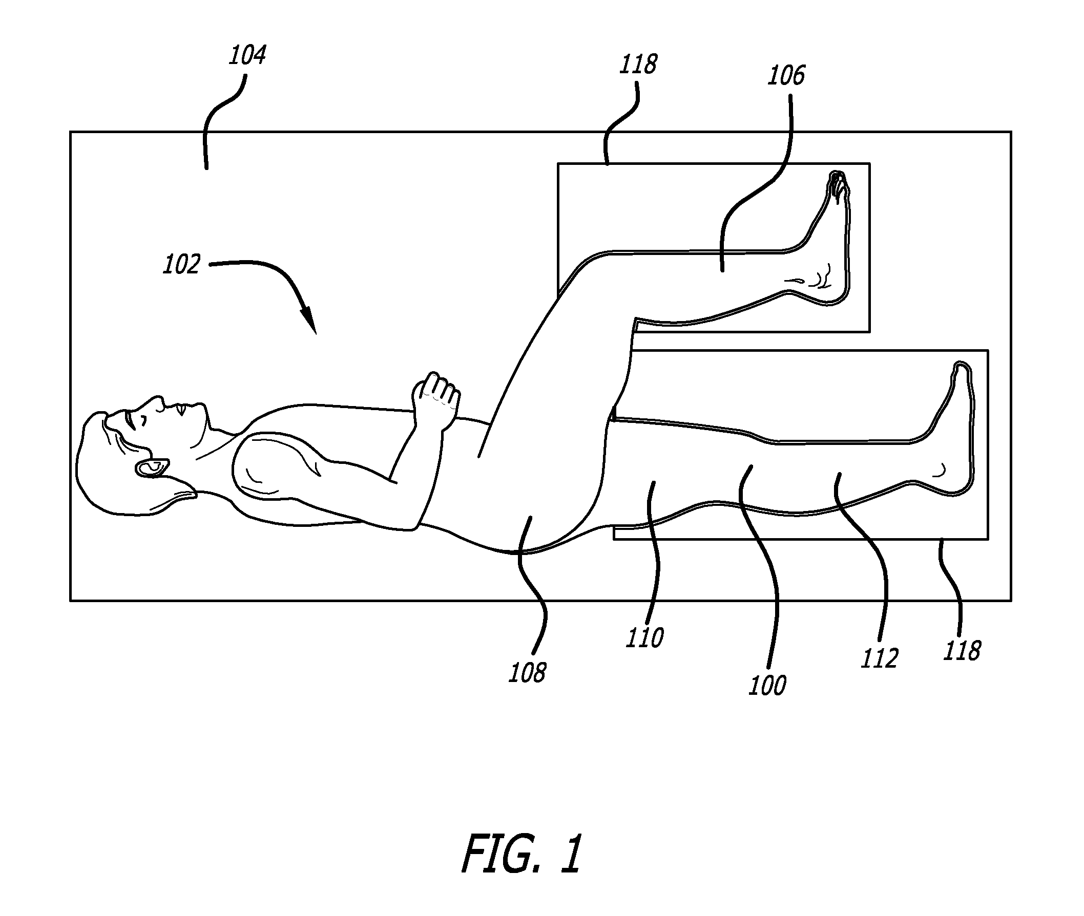
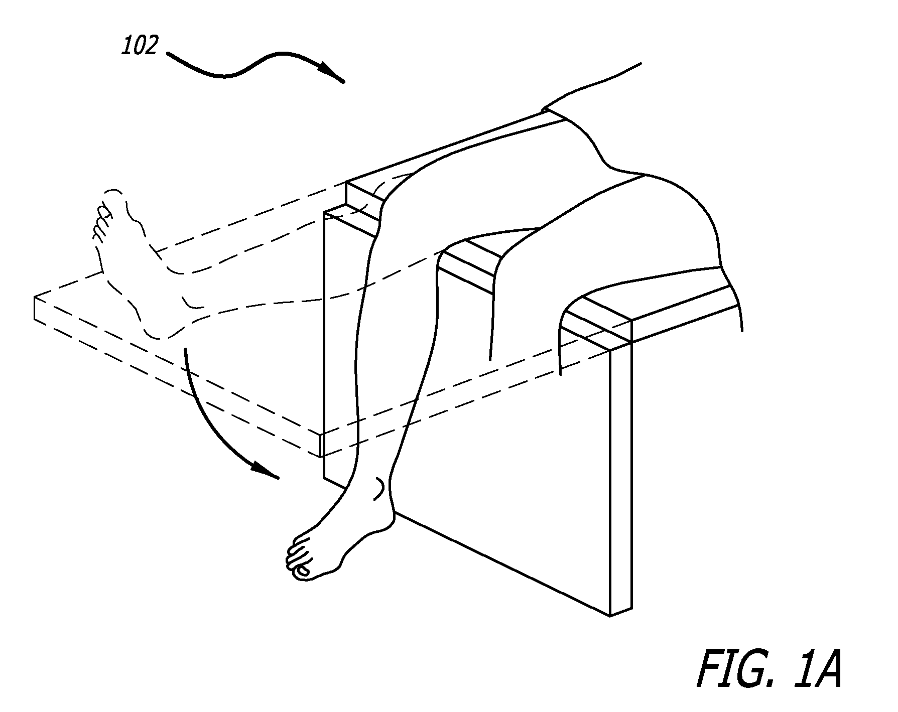
[0035]In one contemplated approach, the position of the patient and treatment areas are selected for easy access and to achieve proper alignment at an implantation site. Use of vacuum lock supports and arch bed structure accomplishes desired treatment site stabilization and orientation. An adjustable surgical table also facilitates desired positioning.
[0037]In further contemplated approaches, proper size and configuration of components of the mechanical energy absorbing apparatus involves understanding the anatomy of the treatment area as well as the unique characteristics of the anatomy of the patient. When the energy absorbing apparatus includes one or more bases which are to be fixed to a bone, such bases are selected to provide surfaces which approximate the bone to which it is to be attached and includes desired separation from the bone to provide connecting structure. In this regard, remote sizing devices and a direct physical inspection of the anatomy is undertaken. Furthermore, fixed distance links, dummy links and base locating tools are employed to facilitate both selection of base implantation sites and proper component identification.
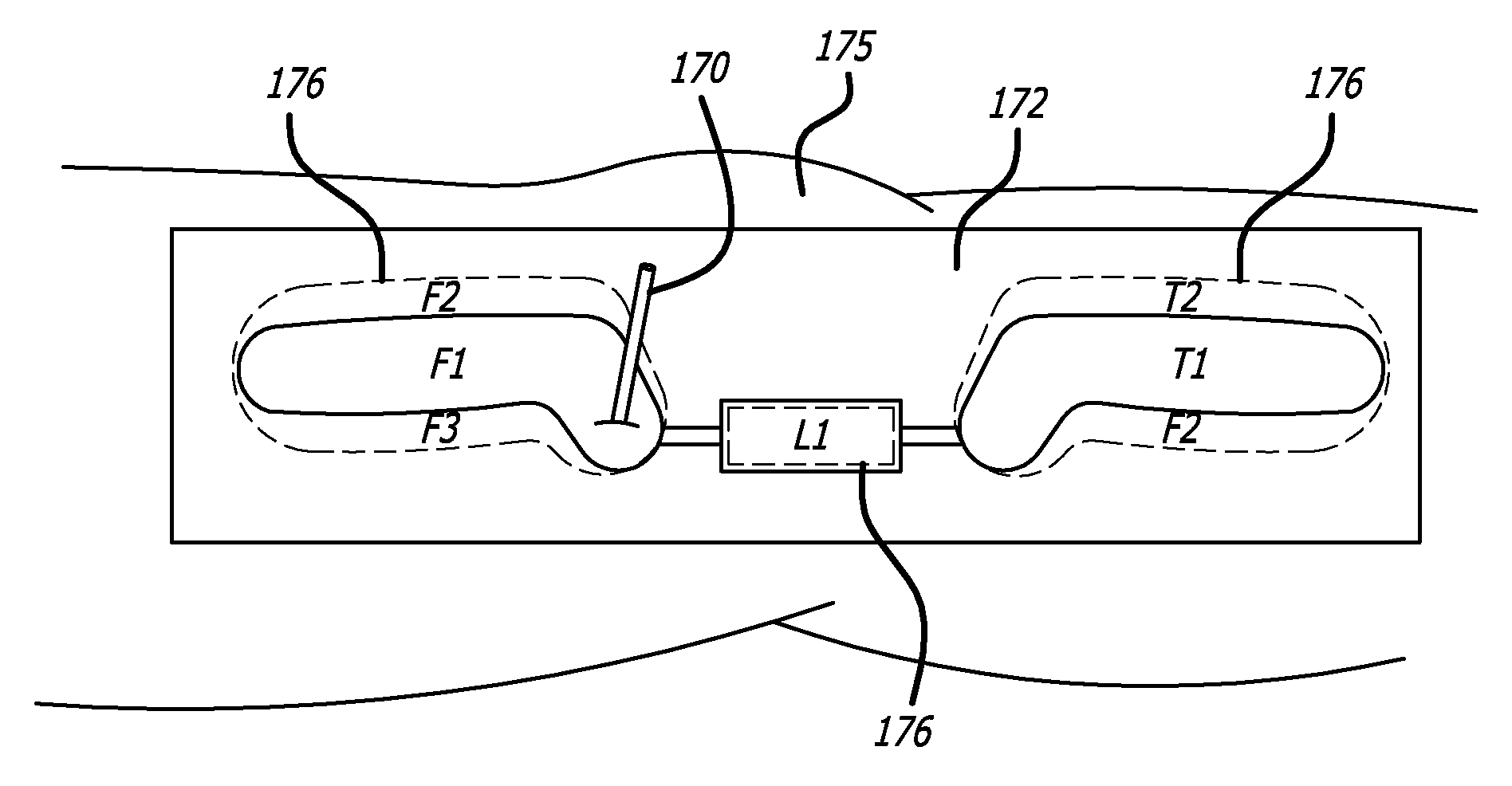
[0041]In one specific embodiment, the presently disclosed apparatus is embodied in a device utilizing an element, or elements functioning as a unit, which responds to bending or changes in elongation. Further, the device is used to reduce the loading experienced by the articular surfaces of the tibiofemoral joint. In one embodiment, the device is designed to reduce load on the joint during knee extension with energy absorption. Joint load reduction in this phase is governed by the compression of the device—increased compression yields greater joint reduction. The device is anchored in a position which ensures device elongation resulting from knee flexion. As the knee moves into flexion, the device is un-compressed and will cause little to no joint load changes. The device may have other features which ensure correct device alignment, and prevent against buckling, as the device transitions into a compressed state. The device can also be configured to provide joint load reductions during flexion or throughout the nearly full range of motion.

Problems solved by technology

Recent experimental studies further indicate that excessive, repetitive loading may induce cell death, and cause morphological and cellular damage, as seen in degenerative joint disease (Lucchinetti et al., 2002 and Sauerland et al., 2003).
Gouty arthritis is caused by deposition of uric acid crystals in the joint that results in subsequent inflammation.
Unfortunately, all arthritides feature pain.
As the bone surfaces become less well protected by cartilage, the patient experiences pain upon weight bearing, including walking and standing.
Due to decreased movement because of the pain, regional muscles may atrophy, and ligaments may become more lax.
The main symptoms of osteoarthritis is chronic pain, causing loss of mobility and often stiffness.
OA can cause a crackling noise (called “crepitus”) when the affected joint is moved or touched, and patients may experience muscle spasm and contractions in the tendons.
Humid weather increases the pain in many patients.
Arthrodeses are described as being reasonable alternatives for treating OA of small hand and foot joints as well as degenerative disorders of the spine, but were deemed to be rarely indicated in large weight-bearing joints such as the knee due to functional impairment of gait, cosmetic problems and further side-effects.
However, it was concluded that such techniques do not presently predictably restore a durable articular surface to an osteoarthritic joint.
Such procedures are a last resort treatment as they are highly invasive and require substantial periods of recovery.
These procedures are also characterized by relatively long recovery times and their highly invasive procedures.
The currently available therapies are not condro-protective.
However, HTO does not address ligamentous instability—only mechanical alignment.
HTO is associated with good early results, but results deteriorate over time.
If a joint surface remains unloaded for appreciable periods of time the cartilage tends to soften and weaken.
Further, as with most materials that experience structural loads, particularly cyclic structural loads, both bone and cartilage begin to show signs of failure at loads that are below their ultimate strength.
There is also a level of load at which the skeleton will fail catastrophically.
Accordingly, it has been concluded that the treatment of osteoarthritis and other conditions is severely hampered when a surgeon is not able to precisely control and prescribe the levels of joint load.
Various of these approaches have had some success in alleviating pain but suffer from patient compliance or lack an ability to facilitate and support the natural motion and function of the diseased joint.
Therefore, mechanical approaches to treating osteoarthritis have had limited applications.
That is, in addition to addressing loads at a joint and joint movement, there has not been an approach which also acknowledges the dampening and energy absorption functions of the anatomy, and taking a minimally invasive approach in implementing solutions.

Method used


Image

  • Surgical implantation method and devices for an extra-articular mechanical energy absorbing apparatus
  • Surgical implantation method and devices for an extra-articular mechanical energy absorbing apparatus
  • Surgical implantation method and devices for an extra-articular mechanical energy absorbing apparatus

Examples


Embodiment Construction

[0129]Referring now to the drawings, which are provided by way of example and not limitation, the present disclosure is directed towards apparatus for treating body tissues. In applications relating to the treatment of body joints, the described approach seeks to alleviate pain associated with the function of diseased or malaligned members forming a body joint. Whereas the present invention is particularly suited to address issues associated with osteoarthritis, the energy manipulation accomplished by the present invention lends itself well to broader applications. Moreover, the present invention is particularly suited to treating synovial joints such as the knee, finger, wrist, ankle and shoulder.

[0130]In one particular aspect, the presently disclosed method seeks to permit and complement the unique articulating motion of the members defining a body joint of a patient while simultaneously manipulating energy being experienced by both cartilage and osseous tissue (cancellous and cor...


PUM


Abstract

A surgical implantation approach for preparing a patient and precisely and effectively placing an energy absorbing apparatus relative to the patient's anatomy. Various surgical implantation apparatus and methods for achieving proper device-to-anatomy juxtapositional relationships are employed in the implantation approach.

Description

CROSS REFERENCE TO RELATED APPLICATIONS[0001]This application is a continuation-in-part of U.S. application Ser. No. 11 / 775,139, filed Jul. 9, 2007, a continuation-in-part of U.S. application Ser. No. 11 / 775,149, filed Jul. 9, 2007 and a continuation-in-part of U.S. application Ser. No. 11 / 775,145, filed Jul. 9, 2007, the entire disclosures of which are expressly incorporated herein by reference.BACKGROUND[0002]The present disclosure is directed towards methods for treating tissue of a body and more particularly, towards procedures and devices for implanting an extra-articular mechanical energy absorbing apparatus.[0003]Both humans and other mammals belong to the subphylum known as vertebrata. The defining characteristic of a vertebrate is considered the backbone or spinal cord, a brain case, and an internal skeleton. In biology, the skeleton or skeletal system is the biological system providing physical support in living organisms. Skeletal systems are commonly divided into three t...

Claims


Application Information

Patent Timeline
IPC IPC(8): A61F2/08
CPCA61B2017/567A61B17/88A61B17/6425A61B17/1739A61B17/1728A61B17/68A61N1/36071A61B17/1764A61F2/3836A61F2/08A61B2019/461A61B2090/061
Inventor CLIFFORD, ANTON G.O'CONNELL, MARYLANDRY, MICHAEL E.
Owner MOXIMED INC
Learn More