Bioactive bone graft substitute
a bone graft and bioactive technology, applied in the direction of prosthesis, drug composition, peptide/protein ingredient, etc., can solve the problems of pain and morbidity, exposing the patient to a second surgery, and limiting the device in terms of shape and size variation
- Summary
- Abstract
- Description
- Claims
- Application Information
AI Technical Summary
Problems solved by technology
Method used
Image
Examples
example 1
Wettability
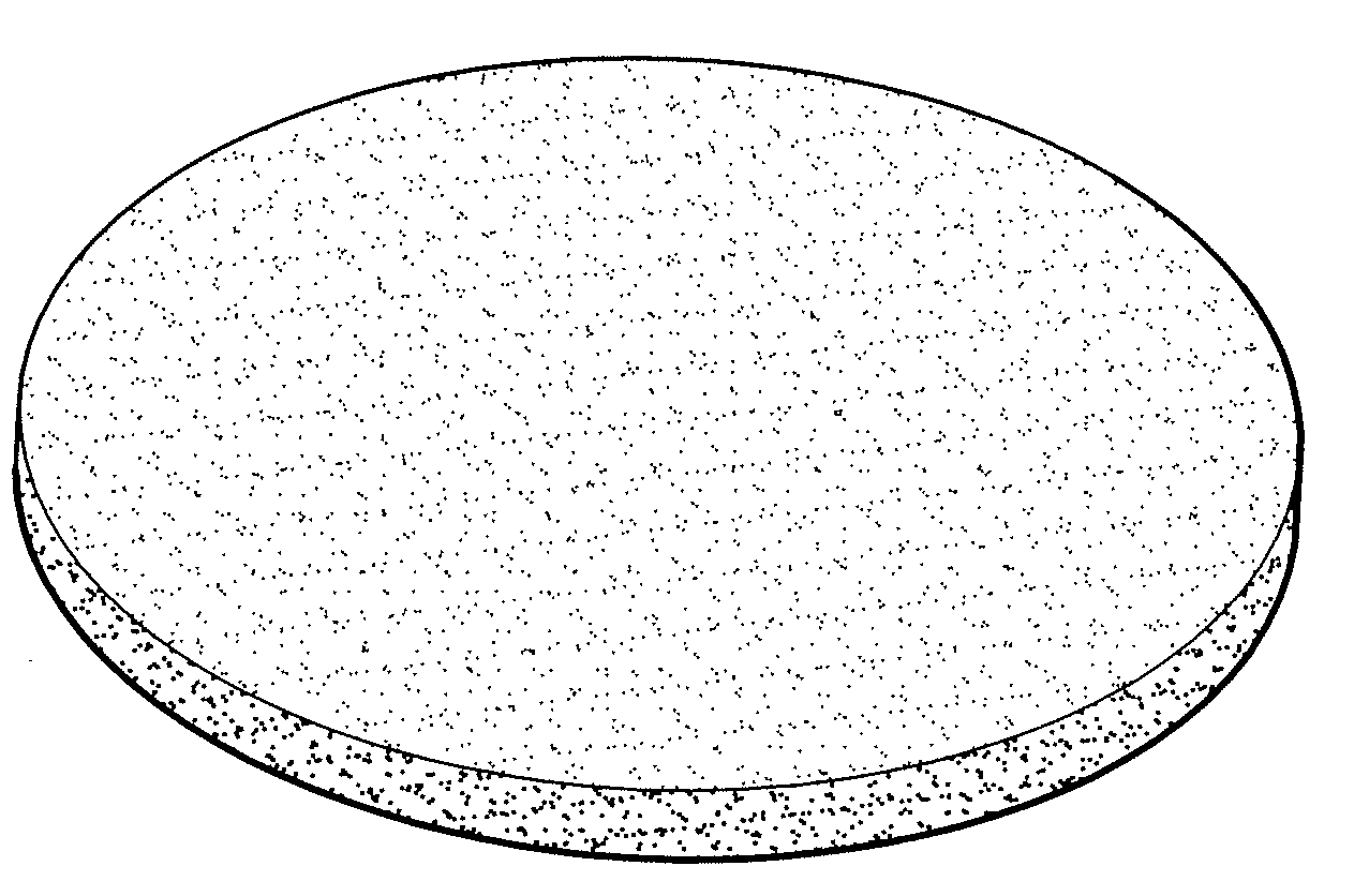
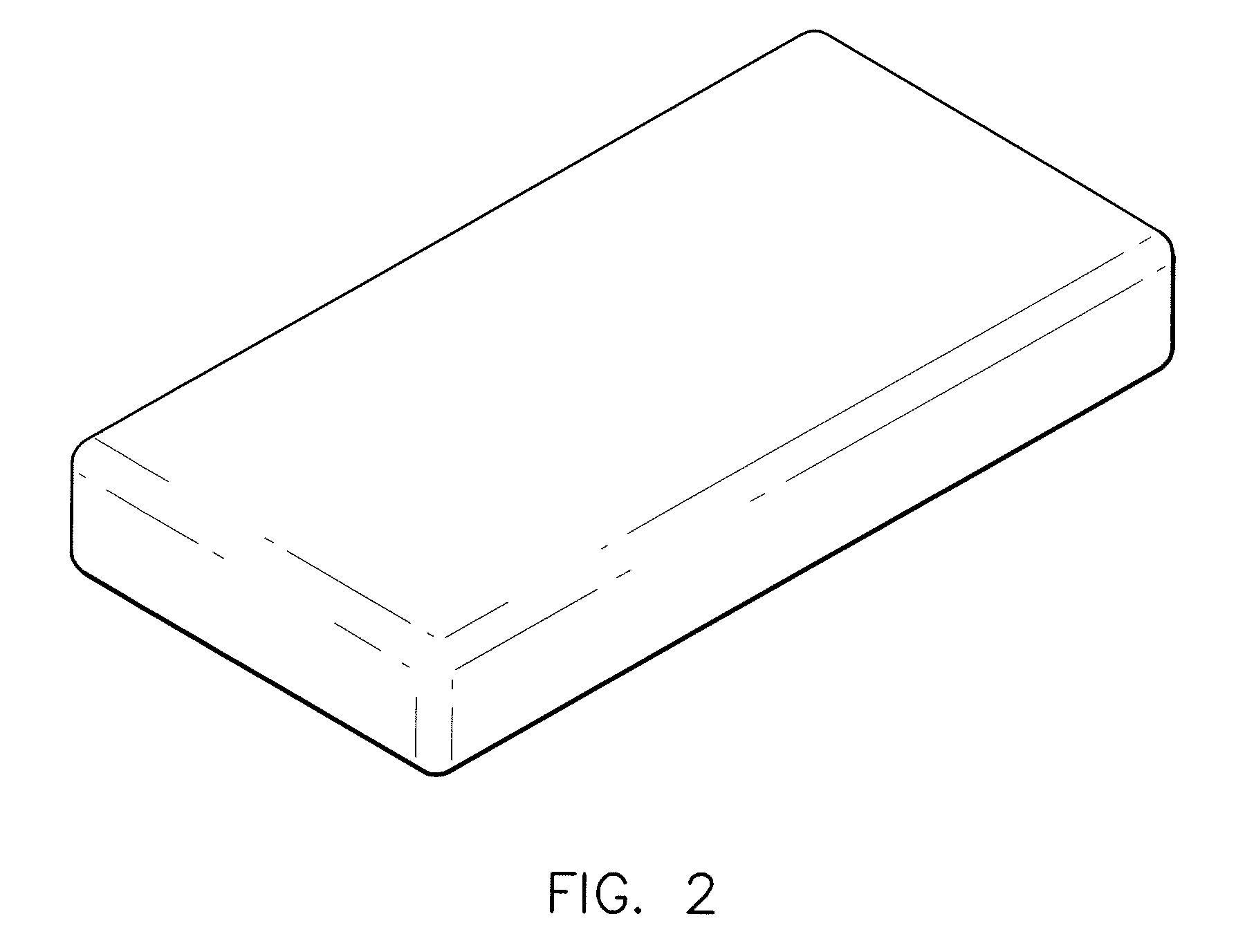
[0094]Dry test samples measuring 25×100×4 mm were weighed and then dipped (“soaked”) in a saline solution for 30 seconds. The weight of the soaked sample was measured. The results of these tests are depicted in Table 1.
TABLE 1WetCalcium phosphateDry WeightWeightMassSample No.(Vitoss ®):Combeite g-c:Collagen(g)(g)Δ (g)IncreaseTest Sample 180:5:154.448613.9679.5184214%Test Sample 280:10:105.80414.5268.722150%Test Sample 380:5:153.39112.449.049267%Test Sample 480:10:104.3313.5899.259214%Control100:0:04.674214.0529.3778201%
example 2
Assessment of Bioactivity
[0095]Bioactivity analysis was conducted on scaffold formulations comprising calcium phosphate, collagen, and bioactive glass in weight ratios of 80:10:10 and 80:15:5. In vitro apatite formation was assessed using SBF, comprising the salts of Na2SO4, K2HPO4·3H2O, NaHCO3, CaCl2, MgCl2·6H2O, NaCl, and KCl. These reagents were dissolved in deionized water and buffered to a pH of approximately 7.3 using Tris (hydroxyl-methyl-amino-methane) and hydrochloric acid. The ionic concentration of the resultant solution closely resembles that of human blood plasma.
[0096]A 1×1 cm specimen of each scaffold formulation was immersed in 20 ml of SBF and incubated at 37° C. At specified time points of 1, 2, and 4 weeks the samples were removed from solution, rinsed with distilled water and acetone, and dried in a dessicator. Bioactivity assessment was carried out using scanning electron microscopy (SEM) and energy dispersive spectroscopy (EDS) to identify changes in surface mo...
example 3
Bioactivity of Calcium Phosphate:Collagen:Bioactive Glass Bone Graft Substitutes (80:15:5) at 4 Weeks
[0097]A sample of calcium phosphate, collagen, and bioactive glass (80:15:5) was immersed in SBF as per the methodology described in Example 2 for 4 weeks. After 4 weeks, SEM and EDAX spectra were taken. As seen in FIGS. 15, 16A, and 17A, new calcium phosphate formation can be identified based on its distinct morphology, as compared to the SEMs of samples prior to SBF immersion. Confirmation of new calcium phosphate formation was confirmed by EDAX spectra (FIGS. 16B, 17B-D). FIG. 16B shows the EDAX spectra of the new growth, confirming its calcium phosphate composition. As further demonstrated in FIGS. 17B and 17D, the new growth has a morphology and associated EDAX spectra distinct from the Vitoss. The EDAX spectra shown in FIG. 17C confirms the collagen strands of the base bone graft substitute visible in FIG. 17A.
PUM
| Property | Measurement | Unit |
|---|---|---|
| particle size | aaaaa | aaaaa |
| particle size | aaaaa | aaaaa |
| total porosity | aaaaa | aaaaa |
Abstract
Description
Claims
Application Information
 - R&D
- Intellectual Property
- Life Sciences
- Materials
- Tech Scout
- Unparalleled Data Quality
- Higher Quality Content
- 60% Fewer Hallucinations
Browse by: Latest US Patents, China's latest patents, Technical Efficacy Thesaurus, Application Domain, Technology Topic, Popular Technical Reports.
© 2025 PatSnap. All rights reserved.Legal|Privacy policy|Modern Slavery Act Transparency Statement|Sitemap|About US| Contact US: help@patsnap.com


