
System and method for cell analysis in microscopy

a cell analysis and microscopy technology, applied in image analysis, image enhancement, instruments, etc., can solve the problems of installation and additional hardware equipmen

Active Publication Date: 2008-08-07
SIEMENS HEALTHCARE DIAGNOSTICS INC
View PDF19 Cites 22 Cited by
  • Summary
  • Abstract
  • Description
  • Claims
  • Application Information

AI Technical Summary

Problems solved by technology

Furthermore, many of them require additional hardware equipment and installation, and may be quite expensive.

Method used


Image

  • System and method for cell analysis in microscopy
  • System and method for cell analysis in microscopy
  • System and method for cell analysis in microscopy

Examples


Embodiment Construction

[0013]According to an embodiment of the present disclosure, a system and method perform cell differentiation and segmentation includes. The method can be applied to both 2D and 3D microscopic cell images, and to a variety of cell types. The method can be extended to 4D where time is an additional parameter, such as in live microscopy or acquiring images in time to track the evolution of certain types of cells. The method enables different types of analyses including counting of cells, identifying structures inside the cells (e.g. existence and shape of nucleus), morphometric analysis of cells (e.g., for shape change assessment and shape analysis), cell surface, volume measurements, color change assessment (e.g. in fluorescence imaging), etc.

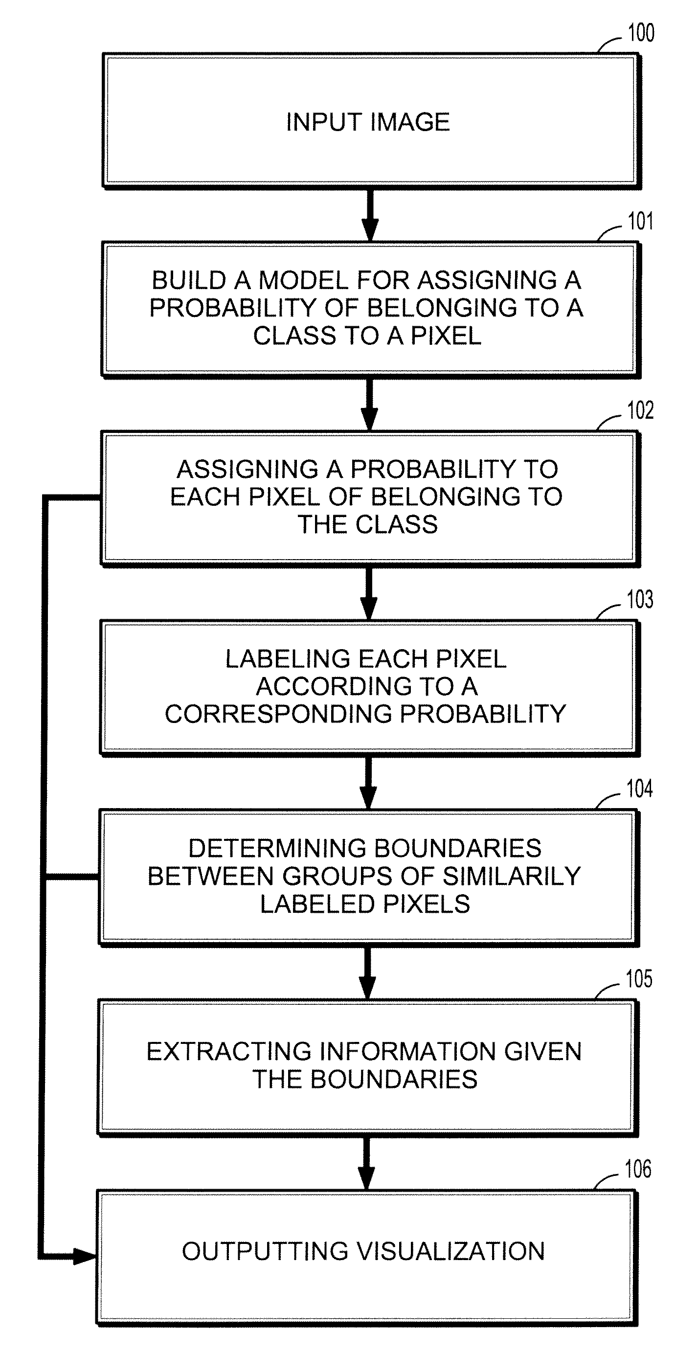
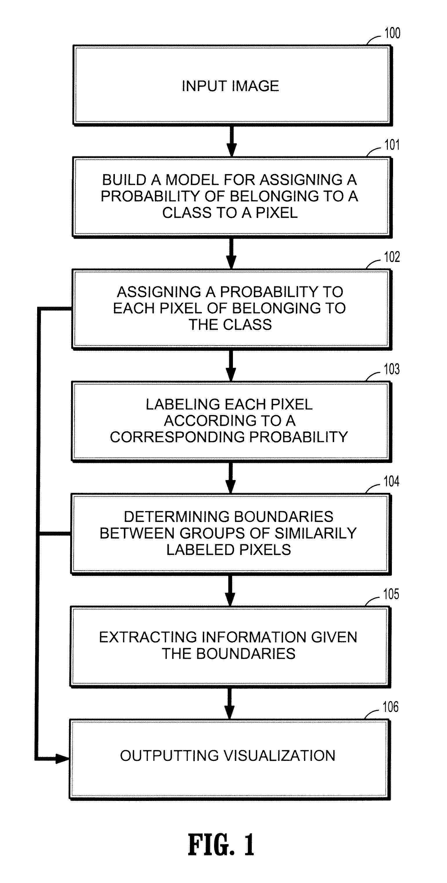
[0014]Referring to FIG. 1, a method for cell differentiation and segmentation includes obtaining a color / intensity model for the cells 101-102, using the model as a set of priors for the random walker segmentation algorithm, segment “cell” pixels...


PUM


Abstract

A computer implemented method for differentiating between elements of an image and a background includes inputting an image comprising pixels forming a view of the elements and a background, providing a model for assigning a probability of belonging to a predefined class to each of the pixels, assigning a probability to each of the pixels of belonging to the predefined class, labeling each of the pixels according to a corresponding probability and a predefined threshold, determining boundaries between groups of like-labeled pixels, and outputting a visualization of the boundaries.

Description

CROSS-REFERENCE TO RELATED APPLICATIONS[0001]This application claims the benefit of Provisional Application No. 60 / 888,147 filed on Feb. 5, 2007 in the United States Patent and Trademark Office, the contents of which are herein incorporated by reference in its entirety.BACKGROUND OF THE INVENTION[0002]1. Field of the Invention[0003]The present disclosure relates to image processing, and more particularly to a system and method for segmenting and counting cells in a microscopic image.[0004]2. Description of Related Art[0005]Images of cells available from 2D and 3D microscopy (and other sources) are increasingly commonplace. For many different applications it is important to analyze the images by segmenting the cells in an image from the background and each other. This segmentation is often a precursor to cell counting or classification (via cell shape, color, etc.) that may be used to answer a variety of diagnostic questions. There are a number of software packages available on the m...

Claims


Application Information

Patent Timeline
IPC IPC(8): G06K9/00
CPCG06K9/0014G06T7/0081G06T7/0093G06T2207/30024G06T2207/20072G06T2207/20101G06T2207/10056G06T7/11G06T7/162G06V20/695
Inventor GRADY, LEOAZAR, FRED S.
Owner SIEMENS HEALTHCARE DIAGNOSTICS INC
Learn More