Device for wound suturing and hemostasis in the thoracic and the abdominal wall mainly in endoscopic operations

a technology for thoracic and abdominal walls, which is applied in the field of endoscopic surgery devices for wound suturing and hemostasis, can solve the problems of high high trauma and labor intensity of methods, and unstable layer structure of wounds, so as to reduce labor and time expenditure, reduce the probability of post-surgery complications, and reduce the traumatizing effect of wound suturing

Inactive Publication Date: 2008-04-24
SHPAICHLER VLADIMIR +1
View PDF14 Cites 35 Cited by
  • Summary
  • Abstract
  • Description
  • Claims
  • Application Information

AI Technical Summary

Benefits of technology

[0021] The object of the present invention is, therefore, the task of developing a device for wound suturing and hemostasis in endoscopic operations that would allow to preserve the initial shape of the wound channel and the stable layer structure of the wound at the moment of suturing thereby diminishing the probability of post-surgery complications. The object of the present invention is further to provide a device that will ensure the initially correct directions of motion of thread-guiding members inserted through the abdominal or the thoracic wall into pre-specified points of the body thereby diminishing the traumatizing effect of wound suturing and reducing labor and time expenditures connected with suturing.

Problems solved by technology

This method is highly traumatizing and labor-consuming as the selection of the right directions of the thread-guiding members inserted through the abdominal or the thoracic wall into the pre-specified points of the body requires quite a considerable amount of time.
Moreover, upon the removal of the port the wound becomes an unstable layer structure.
In its turn, the disarrangement of approximation leads to formation of inter-tissue cavities causing various post-surgery complications such as bleeding, suppuration, and hernias.
The use of this device reduces the traumatizing effect of suturing but layer approximation remains significantly disarranged because of the parallel motion of the threads through the thick sutured tissue.
The use of this device reduces the traumatizing effect during wound suturing but even in this case the wound remains an unstable layer structure, and the approximation of layers of the abdominal wall is disarranged during wound suturing, as described above.
Besides, after the port is removed, the search for the wound channel with the cone apex is often difficult and may cause a change in the wound configuration and a further disarrangement of its layer structure.
Finally, the necessity of searching for the wound channel while using the above devices makes wound suturing after an operation much more difficult.

Method used


Image

  • Device for wound suturing and hemostasis in the thoracic and the abdominal wall mainly in endoscopic operations
  • Device for wound suturing and hemostasis in the thoracic and the abdominal wall mainly in endoscopic operations
  • Device for wound suturing and hemostasis in the thoracic and the abdominal wall mainly in endoscopic operations

Examples


Embodiment Construction

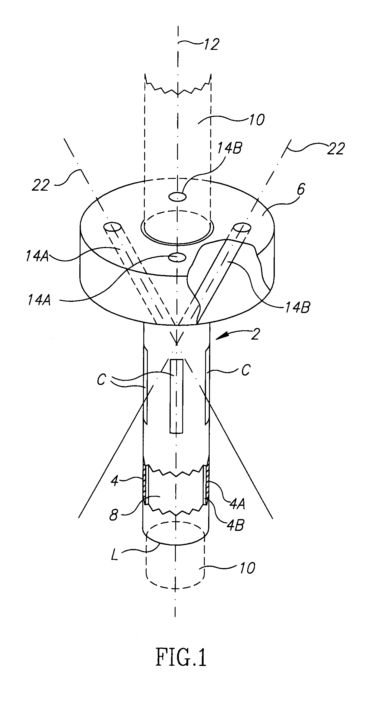
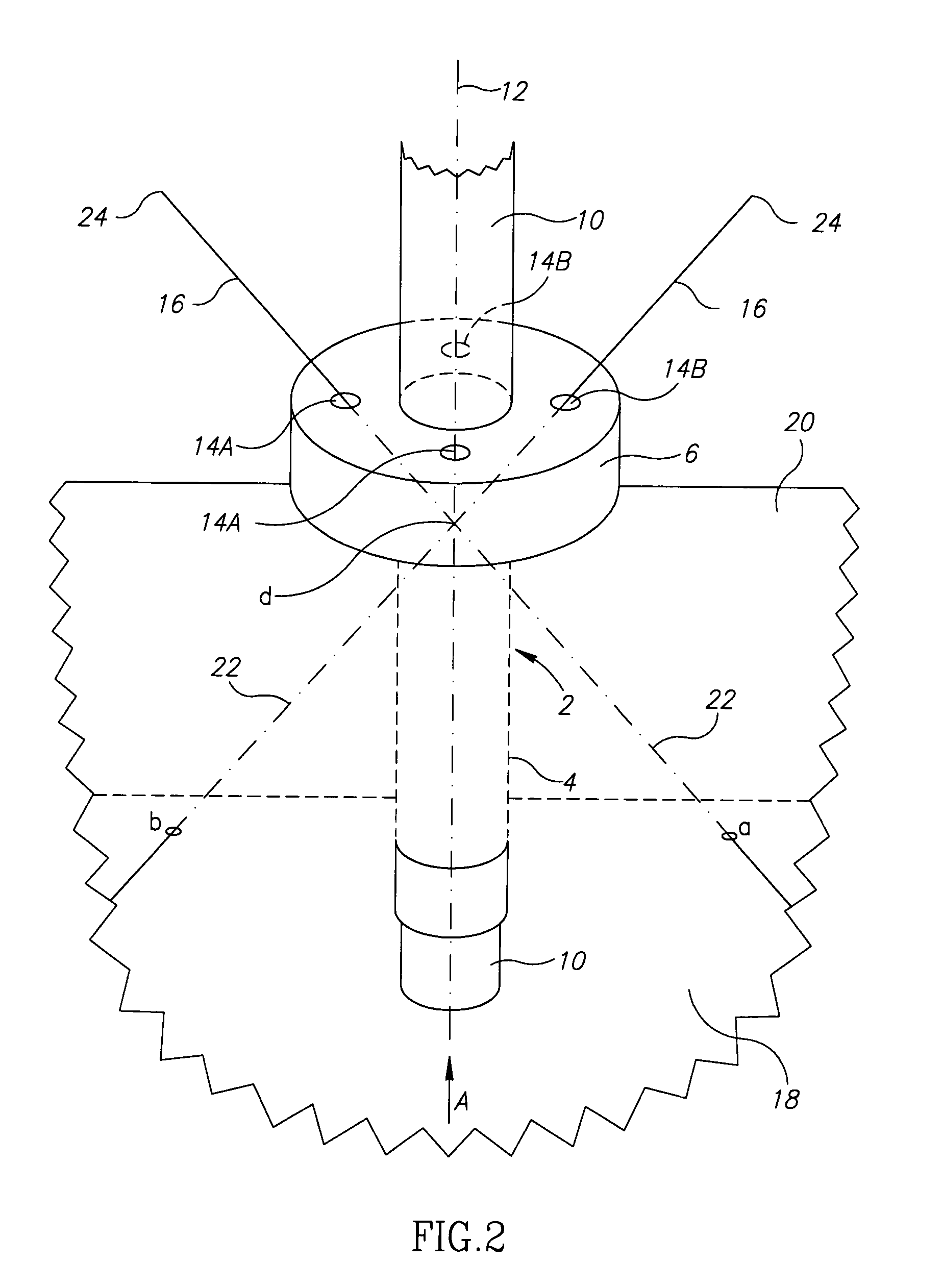
[0030] The proposed device for wound suturing and hemostasis is used after endoscopic operations wherein a port, also at times referred to herein as an endoscopic access port, is employed. The device (FIG. 1)_comprises body 2 having a cylindrical tubular wall 4 equipped with a cap 6 on one end thereof. Tubular wall 4 is intended to preserve the initial shape of wound channel 8 (FIGS. 2,3) during the entire operation. Embodiments of the present invention are possible wherein the tubular wall has other than a cylindrical shape. Outer side 4a of wall 4 in its shape and size (length “L” of the outer cross-sectional perimeter of outer side 4a of wall 4) matches the preset shape and size of wound channel 8. The term “size of wound channel” denotes the cross-sectional perimeter of the wound channel wall. “Size matching” means the equivalence of sizes with acceptable deviations small enough to allow the fulfillment of the object of the present invention. Matching is easy to determine experi...


PUM


Abstract

The aim of the device is to achieve direct and supervised complete closure of the Port Site wound. The following advantages will be procured simultaneously: 1. Preservation of the initial shape of the wound channel. 2. Preservation of the stable layer structure of the Port Site wound during the suturing. 3. Precise, supervised, semi-automatic insertion of the thread-guiding members.

Description

CROSS REFERENCE TO RELATED APPLICATION [0001] This application is a continuation-in-part application of PCT Appl. No. PCT / IL2006 / 000443 (Int. Publ. No. WO 2006 / 111955) filed Apr. 6, 2006, titled “Device for Wound Suturing and Hemostasis in the Thoracic and the Abdominal Wall Mainly in Endoscopic Operations”. The aforementioned PCT application claimed priority from Israel Pat. App. 168129, filed Apr. 19, 2005. FIELD OF THE INVENTION [0002] The present invention relates to the field of medicine and veterinary medicine and, more particularly, to devices for wound suturing and hemostasis in endoscopic, mainly, laparoscopic and thoracoscopic, operations. The invention may also be used for suturing wounds of any etiology similar to those formed during the endoscopic operations. STATE OF THE ART [0003] As it is known, an endoscopic operation comprises the following steps: [0004] insertion of a port through the abdominal or the thoracic wall, into the abdominal or the thoracic cavity, respe...

Claims


Application Information

Patent Timeline
Patent Type & Authority Applications(United States)
IPC IPC(8): A61B17/04
CPCA61B17/3421A61B17/0482
Inventor SHPAICHLER, VLADIMIRSHPAICHLER, EVGENY
Owner SHPAICHLER VLADIMIR
Why Eureka
Try Eureka