Infrared detection of cancerous tumors and other subsurface anomalies in the human breast and in other body parts

Inactive Publication Date: 2007-09-13
BERMAN HERBERT L +2
View PDF1 Cites 100 Cited by
  • Summary
  • Abstract
  • Description
  • Claims
  • Application Information

AI Technical Summary

Benefits of technology

[0020] In a second embodiment, the IR imaging apparatus incorporates a means for manipulating, physically or thermally, the underlying tissue structures utilizing an IR (mid-IR and / or near-IR) transparent window through which at least some of the imaging is performed. Because the “window” is placed against the skin, it can beneficially be used to squeeze or shear the skin or breast tissue as a whole. Squeezing has several benefits including (a) vasculature can be squeezed shut, thereby limiting its thermal contribution, (b) a large area of tissue under investigation is brought in a flat normal incidence angle with the imaging device aiding measurements and removing the angular dependence of emissivity, and (c) if a liquid or gel optical couplant is employed as a thin film between the window and the breast, it can serve to control breast tissue emissivity and emissivity uniformity as well as assure good optical contact. Shearing via rotational or sliding motion of the window drags the surface tissue but “leaves behind” underlying tissue due to tissue shear deformation. Deeper tissues shear less than surface tissues. Thus, by taking images (MIR and / or NIR) at two such sheared positions, one can deduce the depth of the tumor because its surface hotspot moves during the shearing process an amount proportional to its depth.
[0021] In a third embodiment, a preferably IR (mid-IR and / or near-IR) transparent window is utilized as a means of delivering or removing heat from the breast. The window adds or removes heat from the breast either because of its own heat capacity and conductivity or because it includes or is used with a heating or cooling means such as a flowed coolant, electric heater or radiant heater. In any event, heat can be injected or removed in a much more controlled manner and at a much faster and more uniform rate than using prior art cooling means such as blown air or sprayed alcohol. The heat-manipulating window may also be used to thermally cause vasoconstriction in susceptible patients. It may also be used to maintain a large thermal gradient versus tissue depth, thereby further enhancing the thermal contrast of deep tumors. This “heat-plate” or “cold-plate” variation does not require use of an IR transparent window, as it could be used and removed for subsequent imaging. However, we prefer it to be IR transparent and left in place during imaging.

Problems solved by technology

This results in thermal-IR images of breasts that are difficult to interpret correctly, manually or with computer help, in terms of locating the warmer anomalous tumors with certainty.
Using thermal infrared or “thermal-IR” thermography imaging, generally done in the 8-14 micron wavelength regime, it has been demonstrated by several groups over several decades that such underlying tumors have a tissue-surface thermal-IR “heat” signature, albeit that signature is notoriously noisy and currently not alone sufficient to accurately identify such cancers or pre-cancers.
This is not surprising, given modern thermography's sensitivity of better than 0.1° C. However, these thermal images have been noisy in nature and subject to many physiological and environmental factors such that their diagnostic accuracy, in terms of false positives and false negatives, needs improvement.
A physically surface-cooled breast shows enhanced thermal contrast from any embedded heat-producer upon rewarming; unfortunately, these embedded heat-producers also usually include veins and arteries.
Physical cooling of the breast may or may not provide vasoconstricting action.
This works for 80% plus of patients but unfortunately not for all patients.
In some patients, vasoconstriction is not reliable and cannot be used at all
Breast cooling and vasoconstriction with subsequent re-warming and / or removal of vasoconstricting influence help, but not a lot.
The bottom line is that thermography still is not as widely used nor as trusted as is its competitor, the not-hugely better mammography modality.
At this time, the FDA has long-ago approved thermography as an adjunctive to mammography, but thermography does not hold an authoritative clinical position in terms of wide acceptance and reimbursement.
Unfortunately, blown air has poor and non-uniform heat transfer abilities on a three-dimensional breast and sprayed evaporating alcohol is also non-uniform as well as unpleasant if not harmful to skin and respiration.
Mammography itself is far from perfect and probably cannot itself be improved much more beyond making it tomographic rather than 2-D in nature.

Method used


Image

  • Infrared detection of cancerous tumors and other subsurface anomalies in the human breast and in other body parts
  • Infrared detection of cancerous tumors and other subsurface anomalies in the human breast and in other body parts
  • Infrared detection of cancerous tumors and other subsurface anomalies in the human breast and in other body parts

Examples


first main embodiment

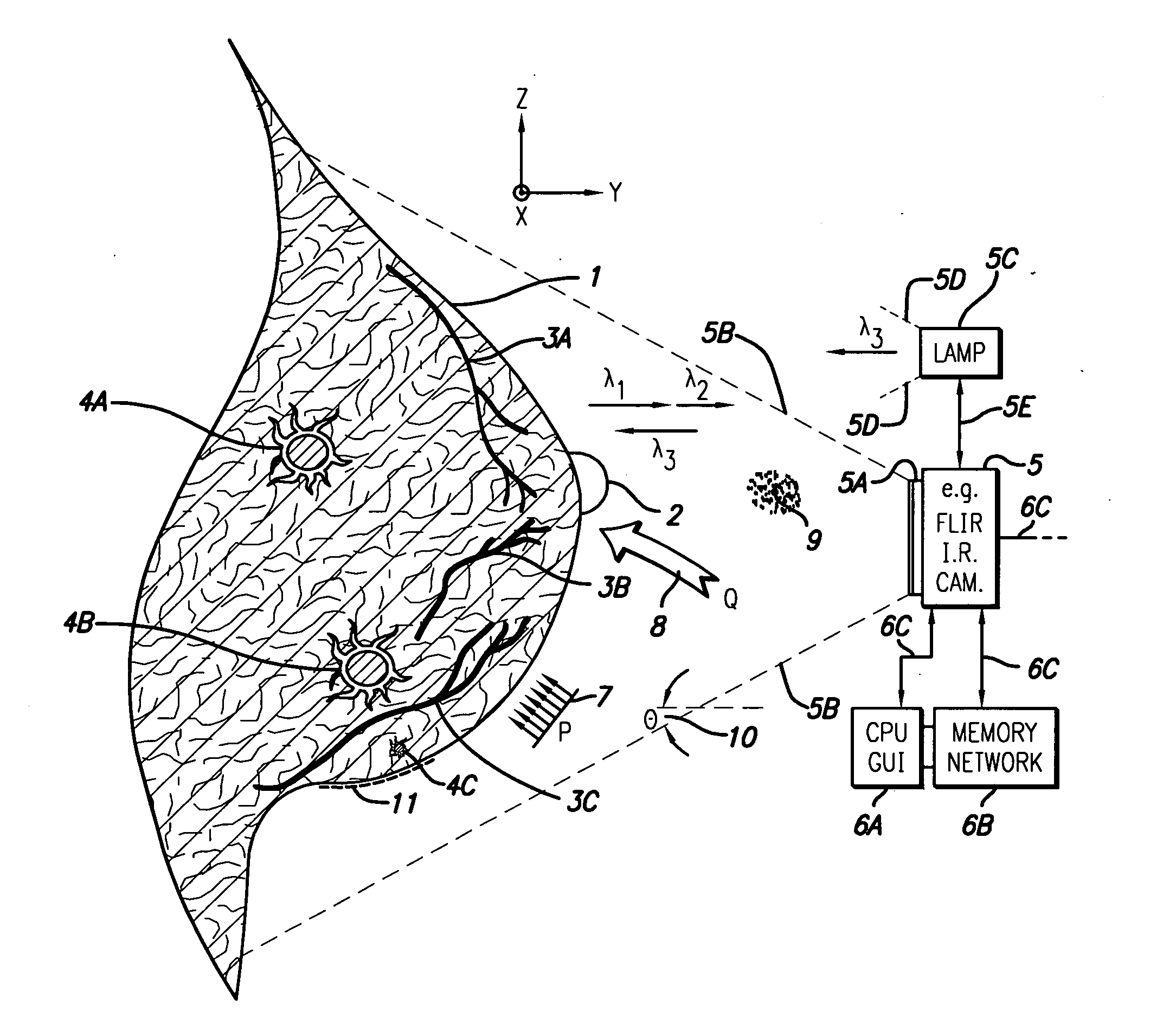
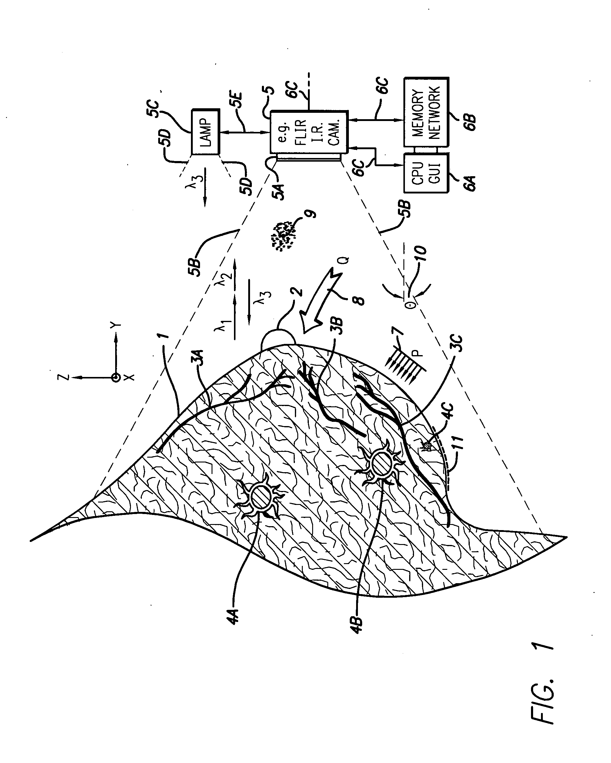
[0063] The first embodiment of the invention utilizes a first nonpenetrating or MIR wavelength to image surface temperatures and a second penetrating wavelength or NIR to image subsurface features. The penetrating or NIR image information is employed in various ways to enhance a determination as to what is diseased and what is not relative to the purely thermographic prior art determination.

[0064] Some ways in which NIR or penetrating wavelength data can do this include the following: [0065] a) using passive or excited near-IR or NIR, delineate the outlines of sub-surface features themselves from at least one point of view and more preferably from two or more points of view, such as of vasculature and / or tumors, without the confounding thermal blooming and masking effects of the mid-IR; [0066] b) using passive or excited near-IR-, delineate any one or more of the size, shape, volume or depth of features themselves from at least one point of view and more preferably from two or more...

second main embodiment

[0077] The second embodiment is essentially the use of a preferably IR (MIR and / or NIR) transmissive or transparent window that can be IR-imaged through while it is also utilized to distort, shear or compress the breast. The preference is to have the window be IR transparent such that IR imaging can be performed while the tissue is distorted. However, within our inventive scope is the use of a plate / window, which is used to distort or compress the breast and is then removed for IR or other imaging without the plate present in the line-of-sight. As described earlier, the window may be flat, curved, rigid or flexible. It may be applied and / or held on the tissue by the clinician's hand, the patient's hand, by straps, clamps or actuator arms, or by suction or adhesive.

[0078] IR transmissive windows, particularly MIR-transparent windows, have been widely used in industrial applications for safety reasons, usually to isolate a worker from high voltages but to still allow MIR visualizatio...

third main embodiment

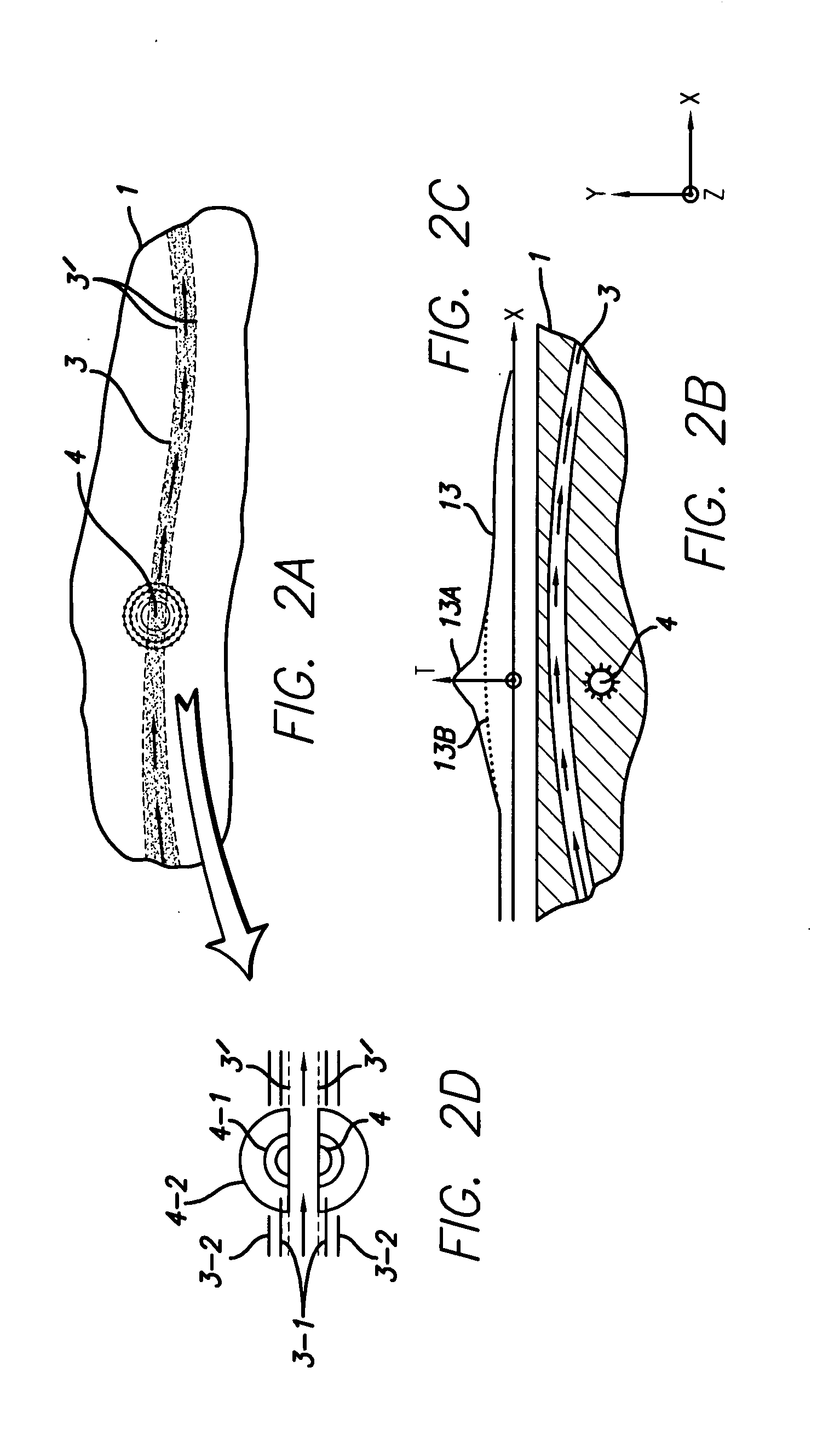
[0115] Moving now to FIGS. 3A-3B, we will describe the use of the second (deforming) plate / window embodiment and the third (heat-manipulating) plate / window embodiment of the invention. The plate / window is a means to thermally and / or physically manipulate tissues while preferably being able to simultaneously observe them at one or more optical wavelengths. The second and third embodiments are not limited to using a penetrating and a non-penetrating wavelength in combination like the first embodiment. Embodiments 2 and 3 may use only one wavelength or may use two or more wavelengths. Embodiments 2 and 3 preferably utilize two or more different states of tissue-deformation and / or tissue-temperature in combination with one (or more) viewing or detection wavelengths.

[0116] A rigid or semi-rigid optical window having a significant thermal capacity, such as our example window materials, can be used to deform tissue and / or inject / remove heat from tissues into or from the window's own therm...


PUM


Abstract

Apparatus and methods to further improve the performance of breast IR-imaging are provided, employing a combination of near-IR and mid-IR frequencies for detection of cancer and other types of subsurface defects. In addition, an IR transmissive or transparent window that can be IR-imaged through is disclosed that may also be utilized to one or both of distort the breast and / or manipulate an artificial heat-flow into or out of the breast.

Description

CROSS-REFERENCE TO RELATED APPLICATION [0001] The present application claims priority from provisional application Ser. No. 60 / 774,562, filed Feb. 16, 2006.BACKGROUND OF THE INVENTION [0002] 1. Field of the Invention [0003] The present invention is directed to the improved detection of cancerous tumors and other subsurface anomalies in human organs or body parts and, in particular, to cancerous growths in the human breast, and, even more particularly, to the use of infrared optical wavelengths for such detection. [0004] 2. Description of Related Art [0005] It has been known from mid-IR thermal-imaging or “thermography” that subsurface breast-cancer tumors present observable infrared thermal (mid-IR) contrast on the external breast tissue surface; however, that contrast is substantially hidden among surface thermal mid-IR contrast or clutter caused by non-anomalous breast vasculature situated at the surface and / or at-depth and having its own thermal mid-IR surface signature. This res...

Claims


Application Information

Patent Timeline
IPC IPC(8): A61B6/00
CPCA61B5/0091A61B5/6843A61B5/4312A61B5/015
Inventor BERMAN, HERBERT L.TOSAYA, CAROL A.SLIWA, JOHN W. JR.
Owner BERMAN HERBERT L
Why Eureka
Try Eureka