
Intubation positioning, breathing facilitator and non-invasive assist ventilation device

a technology of assist ventilation and positioning, which is applied in the field of intubation positioning, breathing facilitator and non-invasive assist ventilation device, can solve the problems of difficult positioning, difficult to adapt the thickness, and do not guarantee always a good visualization, so as to facilitate the spontaneous breathing of obese patients, facilitate the endotracheal intubation, and facilitate the spontaneous inspiration and expiration

Inactive Publication Date: 2007-08-09
MULIER JAN PAUL
View PDF5 Cites 46 Cited by
  • Summary
  • Abstract
  • Description
  • Claims
  • Application Information

AI Technical Summary

Benefits of technology

[0002] The system and method of this invention can be used in three different ways for patients who are lying down in supine position in bed or on the operating table. First it facilitates the endotracheal intubation, secondly it facilitates the spontaneous breathing of obese patients and thirdly it assists the spontaneous inspiration and expiration in a non-invasive way. The sniffing position is the ideal position to visualize the vocal cords during laryngoscopy for intubation of the trachea with an endotracheal tube. The sniffing position consists of a pillow under the head to elevate the head and to improve the alignment of the tracheal axis, pharyngeal axis and axis of the mouth. However these positions do not guarantee always a good visualization, certainly in the obese patients with a short neck where elevation of the upper thorax can help. Instead of elevating the total upper thorax as described until now a folded blanket put under the spinal column of the thorax elevates only the trachea to anterior and stretches the neck.
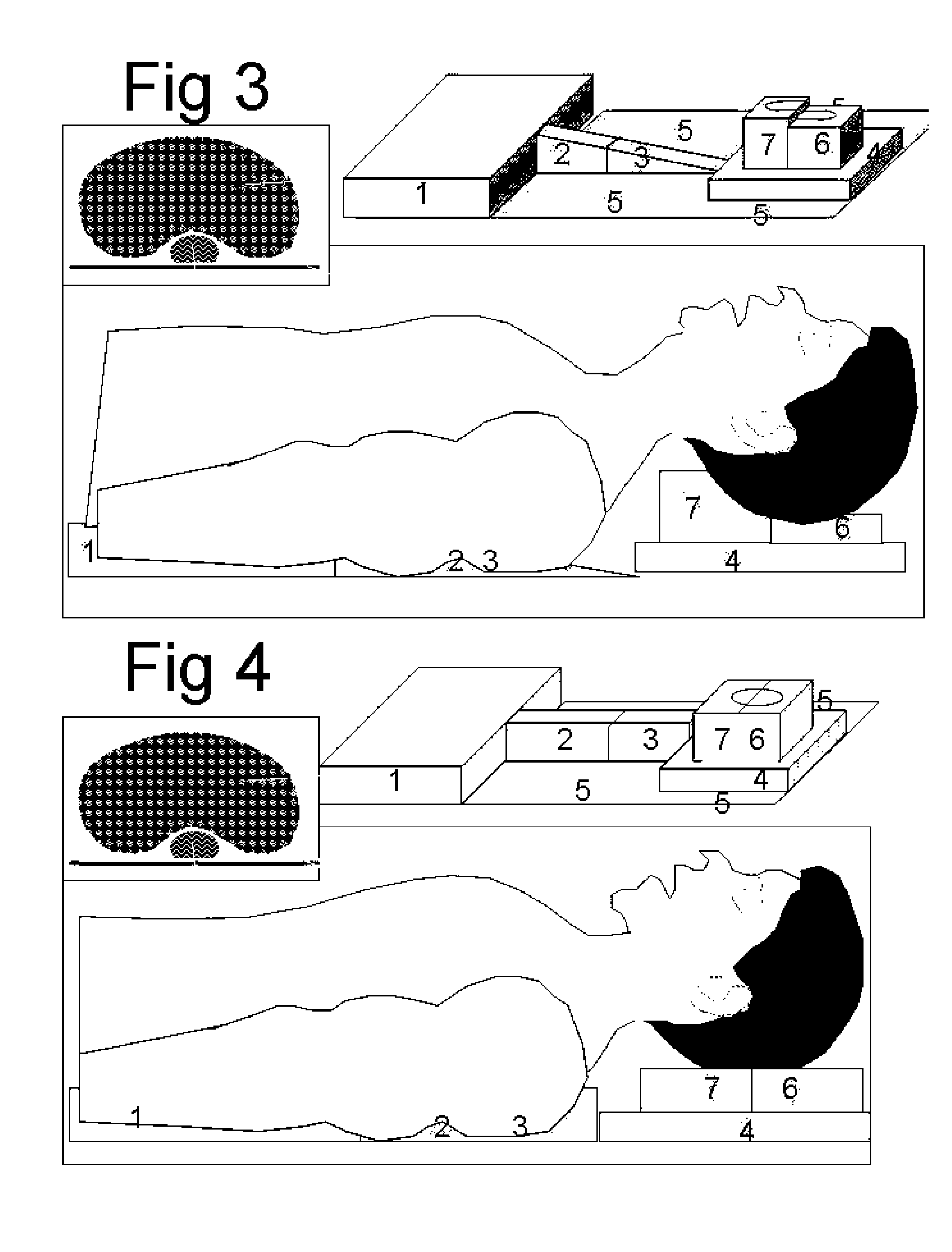
[0003] More exactly the inventor found that elevation of the thoracic spinal column moves the trachea anterior and elevation of the lower thoracic and abdominal spinal column rotates the tracheal axis more in line with the pharyngeal axis. This position together with an elevation and a hyperextension of the head improves further the alignment of the axes and facilitates the laryngoscopy. In most anesthetic courses of intubation however the opposite is learned, because elevation of the upper thorax rotates the trachea opposite and increases the angle between trachea and pharynx, what makes that no one tried to elevate the lower thorax. Certainly in obese patients this helps. It is however difficult to position it before the patient is asleep, difficult to adapt the thickness and difficult to remove after intubation. This invented device is positioned flat under the patient before he is asleep without disturbing him. It allows a gradual elevation and rotation of the spinal column at the desired level of the thorax, a gradual elevation of the head to flex the neck and a gradual hyperextension of the head created by an extension at the atlanto-axial joint. After intubation the position is returned to normal without need of removing the invented device. The device has extra inflatable compartments to elevate the upper thorax if necessary as during trendelenburg position for preventing the patient to slide of the table. A first longitudinal balloon consisting of two compartments, an upper under the upper thorax and a lower under the lower thorax and abdomen is positioned in the length under the spinal column and allows a gradual elevation by inflating both compartments and tilting of the thorax by inflating the lower compartment more than the upper compartment. A second half circular balloon tube under the upper part of the head stabilizes the head and allows a gradual elevation of the head to achieve the sniffing position and a third half circular balloon under the lower part of the head gives a gradual hyperextension of the head. The four gradual movements allow a better ideal alignment of the tracheal axis, the pharyngeal axis and the mouth axis and facilitate intubation. This is the first way to use this invention.
[0004] After the intubation all balloons except the lower under the abdomen are emptied what facilitates central venous puncture if required. During the operation inflation of two triangular balloons under the shoulders, together with an inflation of the head support allow stabilization of the upper thorax and prevention of gliding of the table required in extreme trendelenburg position.
[0005] Spontaneous breathing is easiest when staying upright, certainly when people have to speak, to sing or to blow on an instrument. Total lung capacity decline when a person is lying down and this is certainly true for obese patients. Some persons prefer some extra pillows or want to sleep in a half sitting position. When an obese patient with a hollow back at his thoracic level is lying down, the weight is supported by the ribs of the right and the left side of the thorax and compresses the thorax to a smaller volume. At each inspiration more force is needed to rotate the ribs and to elevate the heavy thorax than when the right and left side of the thorax can move free.
[0006] This invention can elevate the spinal column at the upper and at the lower thoracic level and therefore the thorax is no more compressed and the ribs can move free. Inspiration requires less force and the patient can be breathing easier even when lying down. Obese and non-obese patients can benefit from this support system according to the hollow structure of their back. This is the second way to apply this invention.
[0007] In this invention the upper and lower spinal column elevation system can also be inflated in a synchronized way with the respiration of the patient. During inspiration the spinal column is elevated increasing the lung capacity, increasing the thoracic compliance and facilitating the inspiration. The ribs rotate due to loss of weight comparable with the action of the inspiratory muscles of the thoracic wall. During expiration the elevation is lowered, compressing the thorax, lowering the compliance and therefore facilitating the expiration. The ribs rotate back opposite to the action of the inspiratory muscles. The work of breathing is reduced for the patient resulting in larger minute volume ventilation or less oxygen consumption.

Problems solved by technology

However these positions do not guarantee always a good visualization, certainly in the obese patients with a short neck where elevation of the upper thorax can help.
It is however difficult to position it before the patient is asleep, difficult to adapt the thickness and difficult to remove after intubation.

Method used


Image

  • Intubation positioning, breathing facilitator and non-invasive assist ventilation device
  • Intubation positioning, breathing facilitator and non-invasive assist ventilation device
  • Intubation positioning, breathing facilitator and non-invasive assist ventilation device

Examples


Embodiment Construction

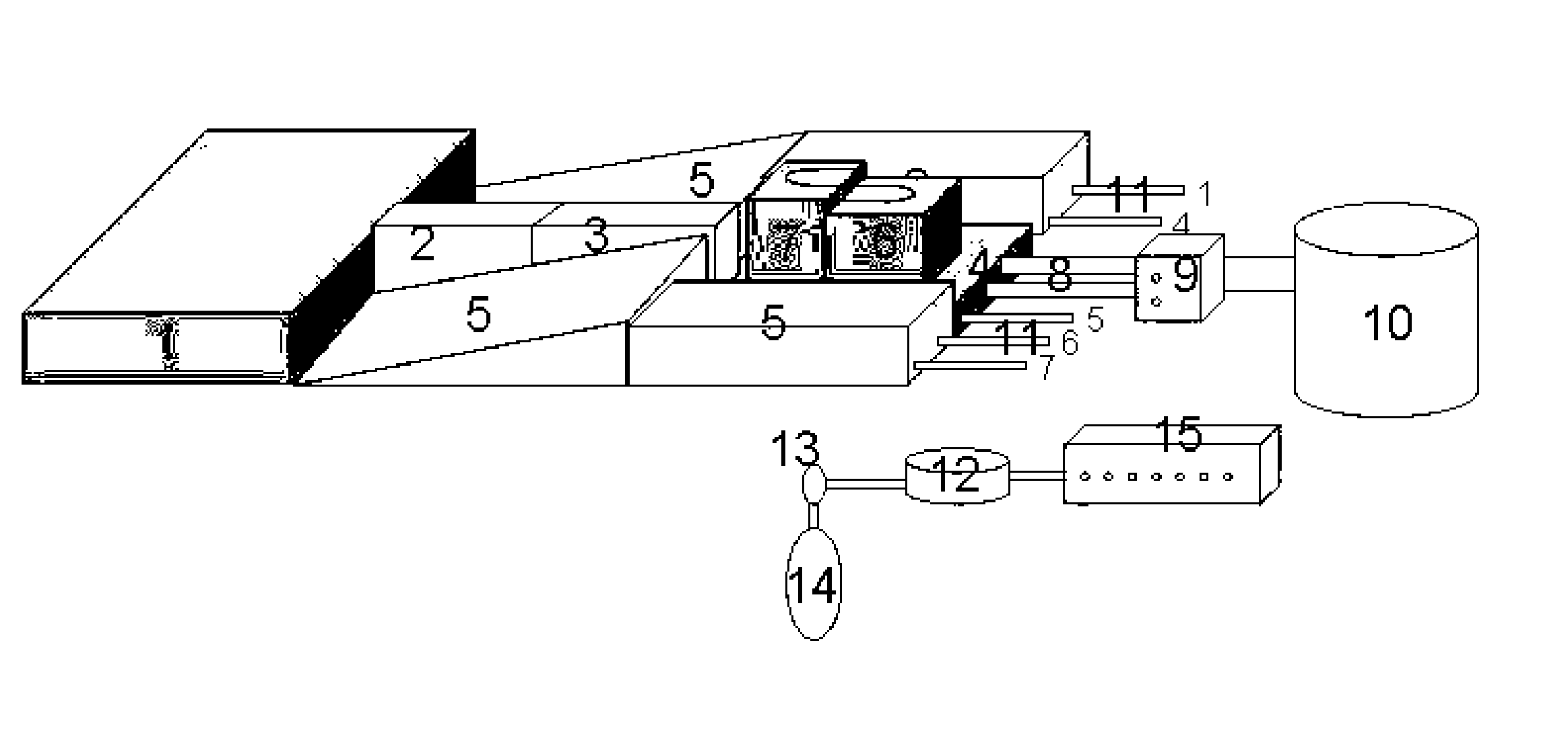
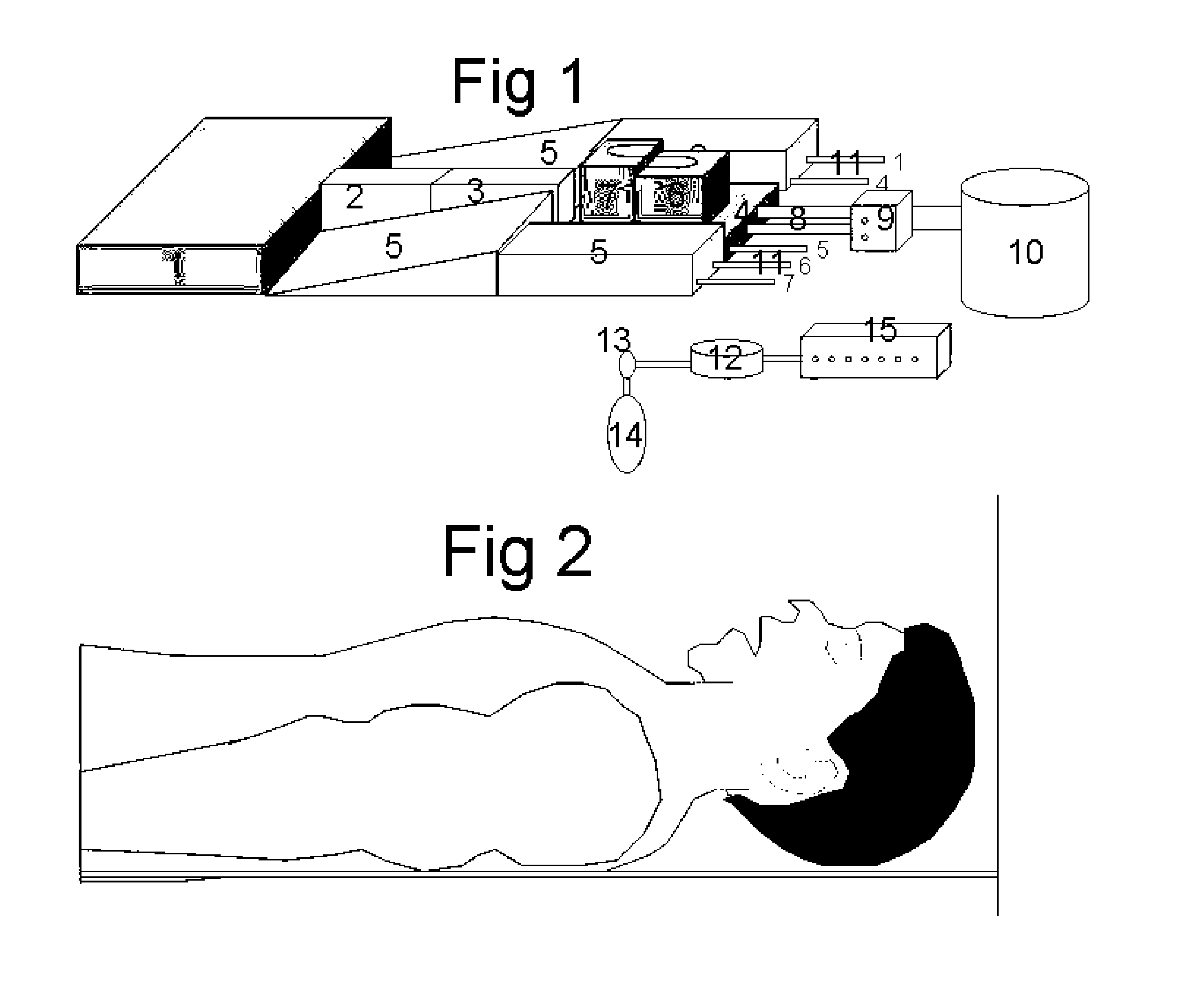
[0015] An intubation positioning, breathing facilitator and non-invasive assist ventilator device in accordance with a preferred embodiment of the present invention, is shown in FIG. 1 where all the balloons of the device are inflated. Number 1 is a flat balloon pillow positioned under the abdomen. Number 2 is a tube like balloon under the lower thoracic spinal column. Number 3 is the balloon positioned under the upper thoracic spinal column needed to inclinate the trachea. Number 4 is a triangular balloon on the right and on the left side positioned under the shoulders. Number 5 is a flat balloon pillow positioned under the head. Number 6 is a half circular balloon positioned under the upper head to elevate the head. Number 7 is a second half circular balloon positioned under the lower head to allow hyper extension of the head during intubation. Number 8 is a double large tube connection, one to balloon number 2 and one to balloon number 3 to allow rapid inflation and deflation whe...


PUM


Abstract

This invention can be used in three different ways for patients who are lying down in bed or on the operating table. First it facilitates the endotracheal intubation, secondly it facilitates the spontaneous breathing of obese patients and thirdly it assists the spontaneous inspiration and expiration in a non-invasive way. This invention device is positioned under the patient before he is asleep without disturbing him. It allows a gradual elevation of the lower and or upper thorax, a gradual elevation of the head giving a flexion of the neck and a gradual hyperextension of the head. After intubation the position is returned to normal without need for removing the invention device. This invention elevates the spinal column and therefore the thorax is no more compressed and the ribs can move free. Inspiration requires less force and the patient can be breathing easier even when lying down. In this invention the spinal column elevation can also be inflated in a synchronized way with the respiration of the patient. During inspiration the spinal column is elevated, facilitating the inspiration. During expiration the elevation is lowered, facilitating the expiration. The work of breathing is reduced for the patient resulting in larger minute volume ventilation or less oxygen consumption.

Description

FIELD OF THE INVENTION [0001] The present invention relates to a system and method that facilitates the endotracheal intubation and the spontaneous breathing of patients and that stimulates the spontaneous breathing of patients in a non-invasive way. In most patients only one of the three applications can be used, although some patients might benefit from more than one application. SUMMARY OF THE INVENTION [0002] The system and method of this invention can be used in three different ways for patients who are lying down in supine position in bed or on the operating table. First it facilitates the endotracheal intubation, secondly it facilitates the spontaneous breathing of obese patients and thirdly it assists the spontaneous inspiration and expiration in a non-invasive way. The sniffing position is the ideal position to visualize the vocal cords during laryngoscopy for intubation of the trachea with an endotracheal tube. The sniffing position consists of a pillow under the head to e...

Claims


Application Information

Patent Timeline
Patent Type & Authority Applications(United States)
IPC IPC(8): A61M15/00A61B5/08A62B7/00
CPCA61G13/12A61G13/121A61G13/1215A61M16/0488A61G13/1255A61G13/1265A61G13/1225
Inventor MULIER, JAN PAUL
Owner MULIER JAN PAUL
Learn More