
Method of recognizing abnormal tissue using the detection of early increase in microvascular blood content

a microvascular blood content and abnormal tissue technology, applied in the field of light scattering and absorption, can solve problems such as difficult detection and usability of methods

Inactive Publication Date: 2007-08-02
NORTHSHORE UNIV HEALTHSYST +1
View PDF55 Cites 38 Cited by
  • Summary
  • Abstract
  • Description
  • Claims
  • Application Information

AI Technical Summary

Benefits of technology

[0009] In one aspect, the present invention describes a method of providing an indication that living tissue within an organ of a body may be abnormal that includes identifying tissue of the organ that contains microvasulature therein, wherein the tissue does ...

Problems solved by technology

While such techniques have advantages in and of themselves as compared to other methods, they require testing of the abnormal tissue itself, which may be difficult to detect.
Further, such methods are usable only after the abnormality is sufficiently large, such as a cancerous tissue.

Method used


Image

  • Method of recognizing abnormal tissue using the detection of early increase in microvascular blood content
  • Method of recognizing abnormal tissue using the detection of early increase in microvascular blood content
  • Method of recognizing abnormal tissue using the detection of early increase in microvascular blood content

Examples


examples

[0055] Without intent to limit the scope of the invention, exemplary instruments, apparatus, methods and their related results according to the embodiments of the present invention are given below. Note that titles or subtitles may be used in the examples for convenience of a reader, which in no way should limit the scope of the invention. Moreover, certain theories are proposed and disclosed herein; however, in no way they, whether they are right or wrong, should limit the scope of the invention so long as the invention is practiced according to the invention without regard for any particular theory or scheme of action.

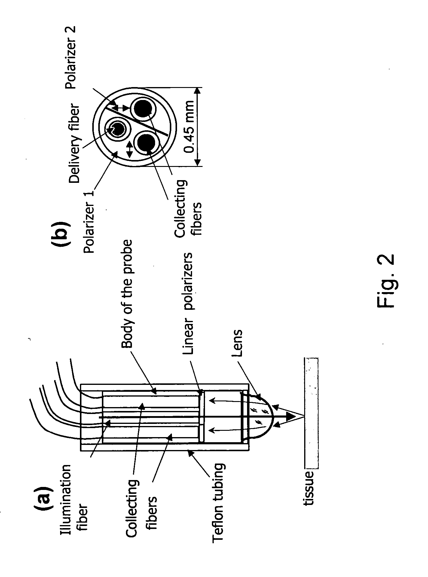
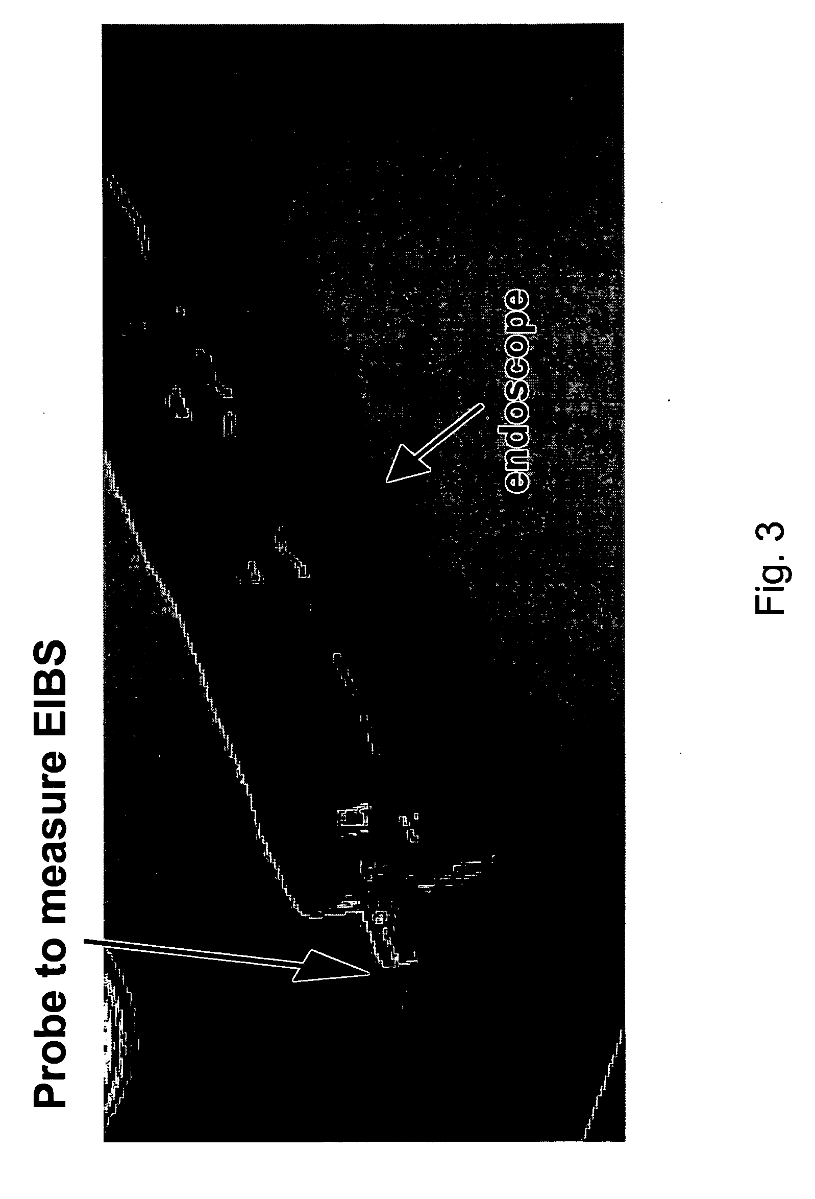
[0056] Polarization Gated Fiber-Optic Probe to Detect EIBS: In one aspect, a fiber-optic probe has been developed to accurately detect blood supply in tissue mucosa. FIG. 2 illustrates the design of the probe in one embodiment and FIG. 3 shows a photograph of the probe protruding from an accessory channel of a colonoscope. The probe has one or more 100 μm-diameter f...


PUM


Abstract

The present invention, in one aspect, relates to a method for examining a target for tumors or lesions using what is referred to as “Early Increase in microvascular Blood Supply” (EIBS) that exists in tissues that are close to, but are not themselves, the abnormal tissue and in tissues that precede the development of such lesions or tumors. While the abnormal tissue can be a lesion or tumor, the abnormal tissue can also be tissue that precedes formation of a lesion or tumor, such as a precancerous adenoma, aberrant crypt foci, tissues that precede the development of dysplastic lesions that themselves do not yet exhibit dysplastic phenotype, and tissues in the vicinity of these lesions or pre-dysplastic tissues.

Description

PRIORITY CLAIM [0001] This application claims priority to U.S. Application No. 60 / 801,947 entitled “Guide-To-Colonoscopy By Optical Detection Of Colonic Micro-Circulation And Applications Of Same”, which was filed on May 19, 2006, the contents of which are expressly incorporated by reference herein. This application is also a continuation-in-part and claims priority to copending U.S. patent application Ser. No. 11 / 261,452 entitled “Multi-Dimensional Elastic Light Scattering”, filed Oct. 27, 2005 with the same assignee as the present invention, the disclosure of which is incorporated in its entirety herein by reference. [0002] Some references, which may include patents, patent applications and various publications, are cited and discussed in the description of this invention The citation and / or discussion of such references is provided merely to clarify the description of the present invention and is not an admission that any such reference is “prior art” to the invention described h...

Claims


Application Information

Patent Timeline
IPC IPC(8): A61B5/00
CPCA61B5/0075A61B5/0084A61B5/1459A61B5/0261A61B5/0091
Inventor BACKMAN, VADIMROY, HEMANTWALI, RAMESHKIM, YOUNG L.SIEGEL, MICHAELTURZHITSKY, VLADIMIR
Owner NORTHSHORE UNIV HEALTHSYST
Learn More