Phase-contrast enhanced computed tomography

Inactive Publication Date: 2005-06-16
VANDERBILT UNIV
View PDF18 Cites 13 Cited by
  • Summary
  • Abstract
  • Description
  • Claims
  • Application Information

AI Technical Summary

Benefits of technology

[0016] In another embodiment of this disclosure, there is provided an apparatus which creates phase-contrasted CT images based on interference effects in which both coherence and imaging require the use of perfect crystals. As used herein, a “perfect crystal” is one in which there are no substantial point, linear, or planar imperfections. Specifically, this embodiment of the present invention provides a monochromatic diffraction computed tomography scanner which allows the user to obtain phase-contrast, x-ray computed tomography images of a sample object. The samples that can be scanned by this CT scanner are unlimited. However, in one embodiment, the sample includes biological soft tissues. This CT scanner may include a polychromatic x-ray source. There is also a power supply source coupled to the polychromatic x-ray source to power the polychromatic x-ray source. There is a first translation stage coupled to the power supply source. There is also a pair of rotatable, interchangeable monochromator crystals in spaced relation with the polychromatic x-ray source. A rotational stage in spaced relation with the monochromator crystals and configured to hold and rotate the sample is also included. There is a rotatable, interchangeable analyzer crystal in spaced relation with the rotational stage. There is also a detector in spaced relation with the analyzer crystal and configured to collect phase contrast x-ray data from the sample. A second translation stage coupled to the detector and a computer coupled to the detector and configured to form a computed tomography image of the sample using the phase-contrast x-ray data also exists. This CT scanner may even provide that the computer controls the power supply source, the pair of rotatable interchangeable monochromator crystals, the first translation stage, the rotational stage, the rotatable interchangeable analyzer crystal and / or the second translation stage. In another aspect of this embodiment of this disclosure, the polychromatic x-ray source may include an x-ray tube, a radioactive source and / or a synchrotron radiation source. The CT scanner may even further provide that the polychromatic x-ray source operates at a range between 20 and 150 kVp. This CT scanner may further provide that the pair of monochromator crystals and the analyzer crystal may be selected from a group of non-perfect crystals. However, in one embodiment, the pair of monochromator crystals and the analyzer crystal may be perfect silicon crystals. Finally, in yet another aspect of, this present disclosure, an existing x-ray source can be interchanged with the polychromatic x-ray source which would enable the existing x-ray source to perform phase-contrast imaging.

Problems solved by technology

Current mammography screening has a very high rate of false positives and false negatives (Fitzgerald, 2001).

Method used


Image

  • Phase-contrast enhanced computed tomography
  • Phase-contrast enhanced computed tomography
  • Phase-contrast enhanced computed tomography

Examples


Embodiment Construction

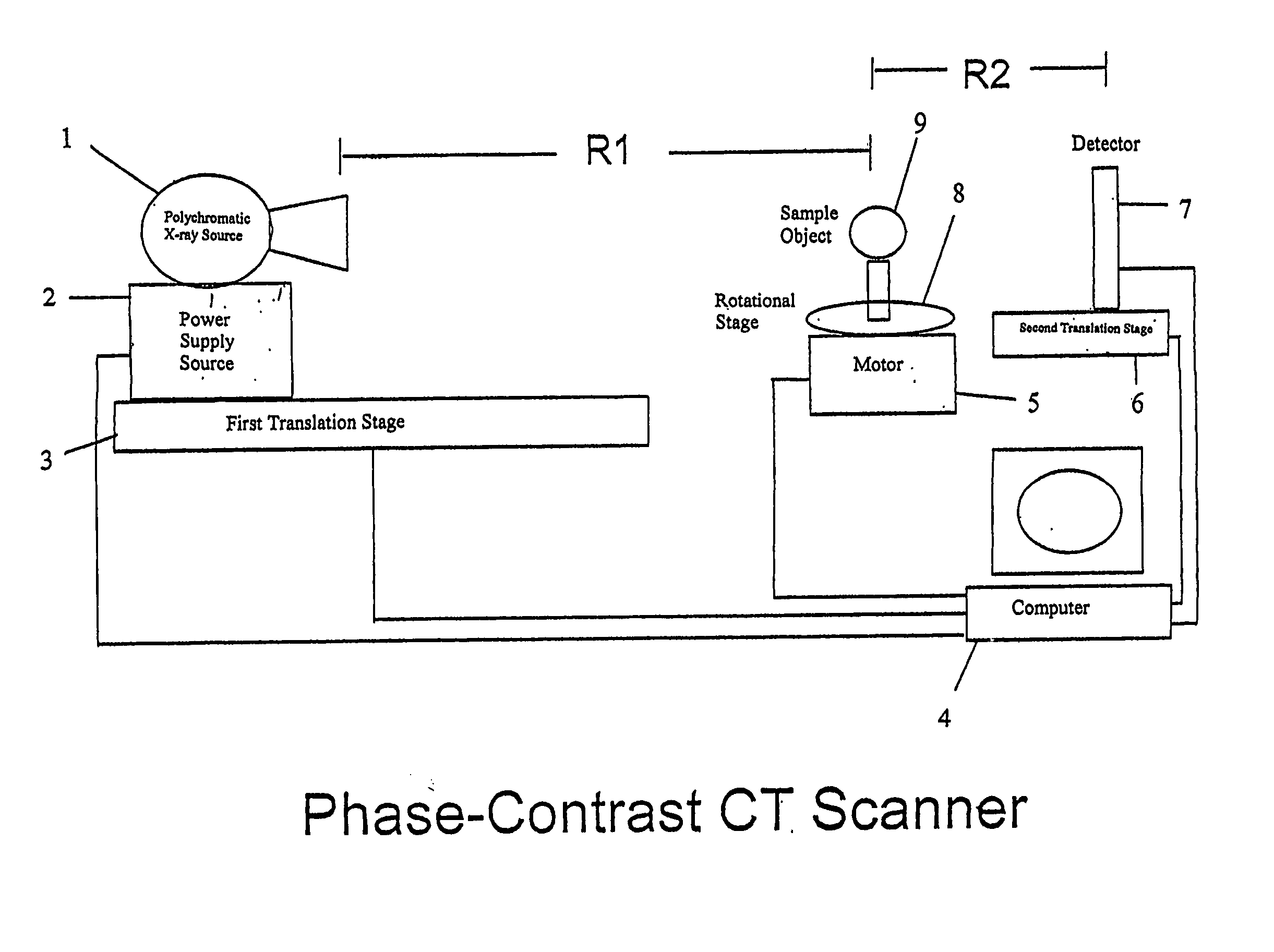
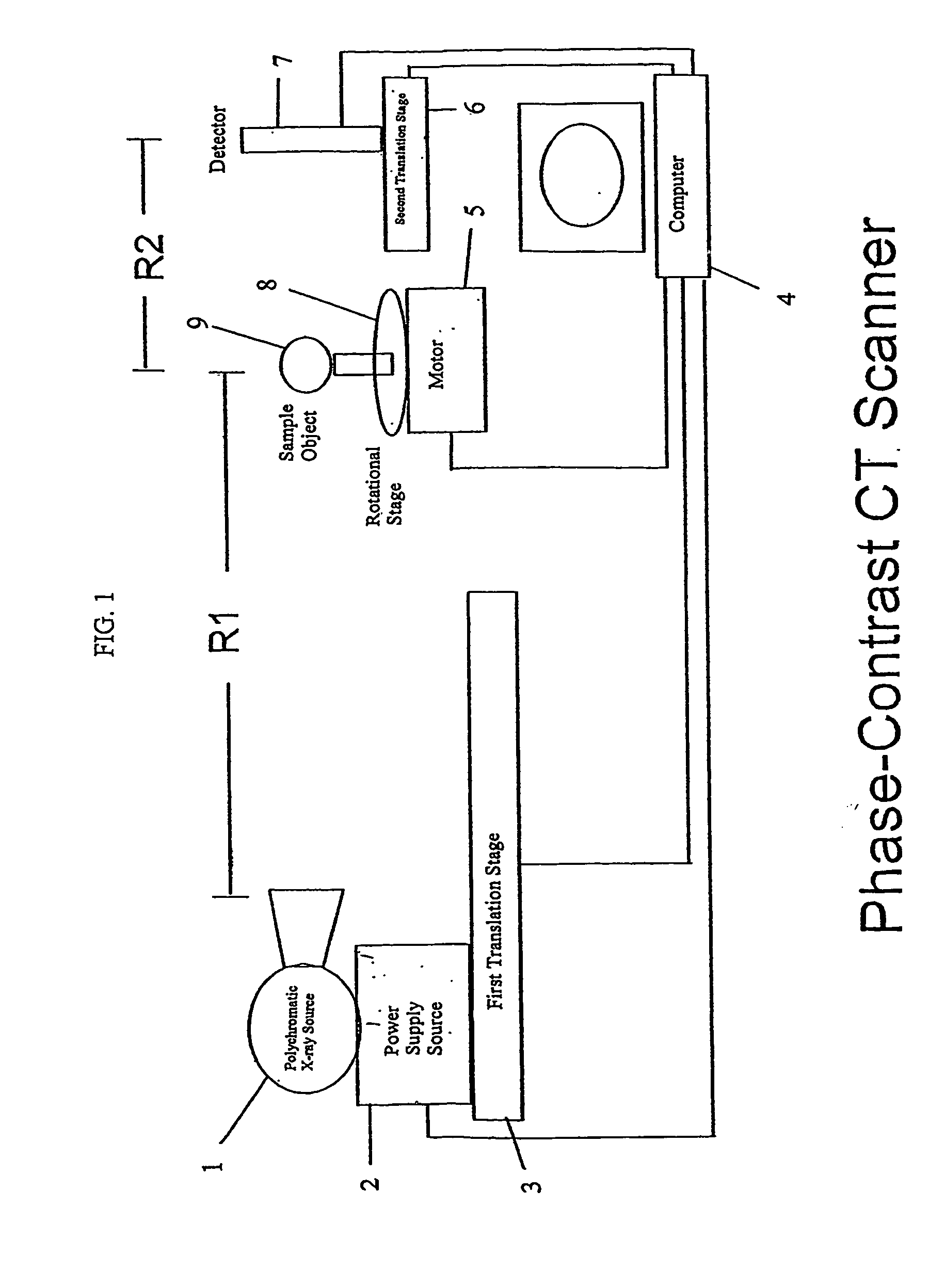
[0024]FIG. 1 illustrates one possible construction of a phase-contrast computed tomography scanner for providing phase-contrast, x-ray computed tomography images of a sample. A polychromatic x-ray source 1 may be connected to and powered by a power supply source 2. In one embodiment, the polychromatic x-ray source 1 may be selected from a group including an x-ray tube, a radioactive source and / or a synchrotron radiation source. The power supply source 2 may operate between a range of 20 and 150 kilovoltage potential. The power supply source 2 may be connected to a computer 4. The computer 4 may control the kilovoltage output of the power supply source 2. In another embodiment, the power supply source 2 may be controlled independent of the computer 4. The polychromatic x-ray source 1 may be situated on top of the power supply source 2. The power supply source 2 may be situated on top of a first translation stage 3. The first translation stage 3 can be maneuvered through an x-axis, a ...


PUM


Abstract

A phase-contrast x-ray computed tomography scanner, a monochromatic diffraction computed tomography scanner, a rotatable monochromatic diffraction computed tomography scanner, and a combination phase-contrast and monochromatic computed tomography scanner are provided. In addition, a method of identifying an unknown sample is provided.

Description

[0001] This application claims priority to, and specifically incorporates herein by reference, U.S. Provisional Application Ser. No. 60 / 338,942 filed Nov. 5, 2001.BACKGROUND OF THE INVENTION [0002] 1. Field of the Invention [0003] The present invention relates generally to the fields of radiography and tomography. More particularly, this disclosure describes how a standard x-ray source may be used to create phase-enhanced computed tomography (CT) images. These phase-enhanced CT images provide sharper images which make small edges, including small masses or small spiculations, more evident. This provides a method for, among other things, the inspection of organic materials and for use in medical apparatuses. [0004] 2. Description of Related Art [0005] In the conventional x-ray transmission imaging system, the contrast of an obtained image depends upon the degree of absorption of x-rays by an object. Namely, if there is a region where heavy elements with high x-ray absorptance are den...

Claims


Application Information

Patent Timeline
IPC IPC(8): G01N23/04
CPCA61B6/484G01N23/046G01N2223/419G21K2207/005A61B6/4092
Inventor DONNELLY, EDWIN F.PRICE, RONALD R.
Owner VANDERBILT UNIV
Why Eureka
Try Eureka