
Endoscope having multiple working segments

a fiberoptic endoscope and working segment technology, applied in the field of flexible fiberoptic endoscopes, can solve the problems of limited scope, large instruments, clumsiness, etc., and achieve the effect of facilitating the performance of multiple-hand surgical procedures

Inactive Publication Date: 2005-04-21
GRANIT MEDICAL INNOVATION
View PDF5 Cites 90 Cited by
  • Summary
  • Abstract
  • Description
  • Claims
  • Application Information

AI Technical Summary

Benefits of technology

"The present invention is an improved fiberoptic endoscope that allows for multiple-hand surgical procedures. It is composed of two or more elongate flexible distal segments joined at a position proximal to the distal end. Each segment may have light and visualization components along with working channels to facilitate medical procedures. The endoscope is designed to be easily inserted into a patient's body cavity and can be joined together to maneuver the working segments independently. The invention also provides a flexible endoscope with a separate segment containing the light and visualization components, which can be joined together to allow for the performance of complex surgical procedures. The endoscope can be controlled by a single operator or two operators in conjunction with an assistant or another endoscopist. The invention expands the scope of therapeutic flexible fiberoptic endoscopy and allows for the performance of surgery within the lumen instead of entering the hollow organ through the abdominal or chest cavity."

Problems solved by technology

Certain minimal surgical procedures are being performed, but these are quite limited in scope.
These instruments are large and clumsy and are, for the most part, attached to the endoscope rather than manipulated through its working channels.

Method used


Image

  • Endoscope having multiple working segments
  • Endoscope having multiple working segments
  • Endoscope having multiple working segments

Examples


Embodiment Construction

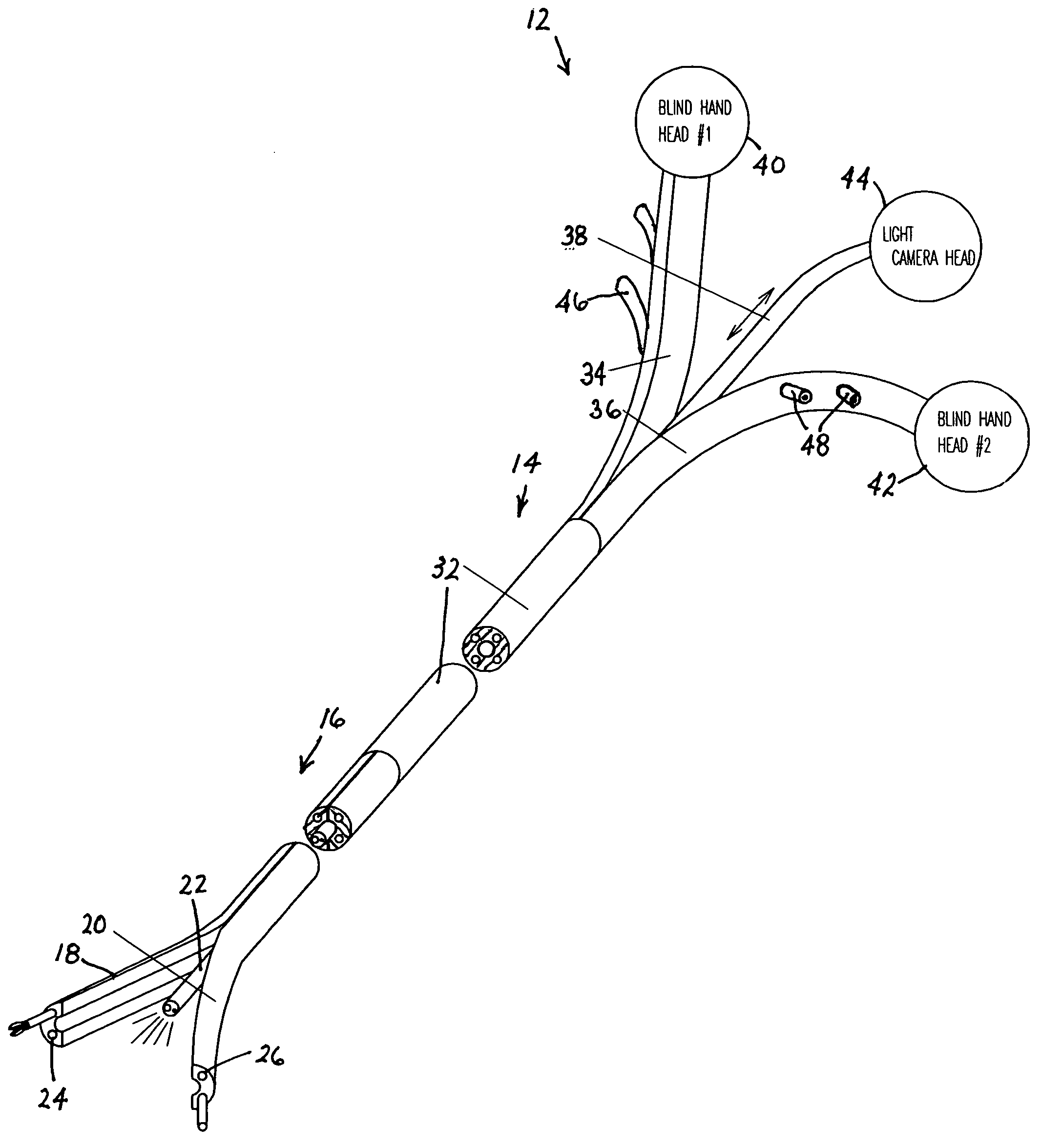
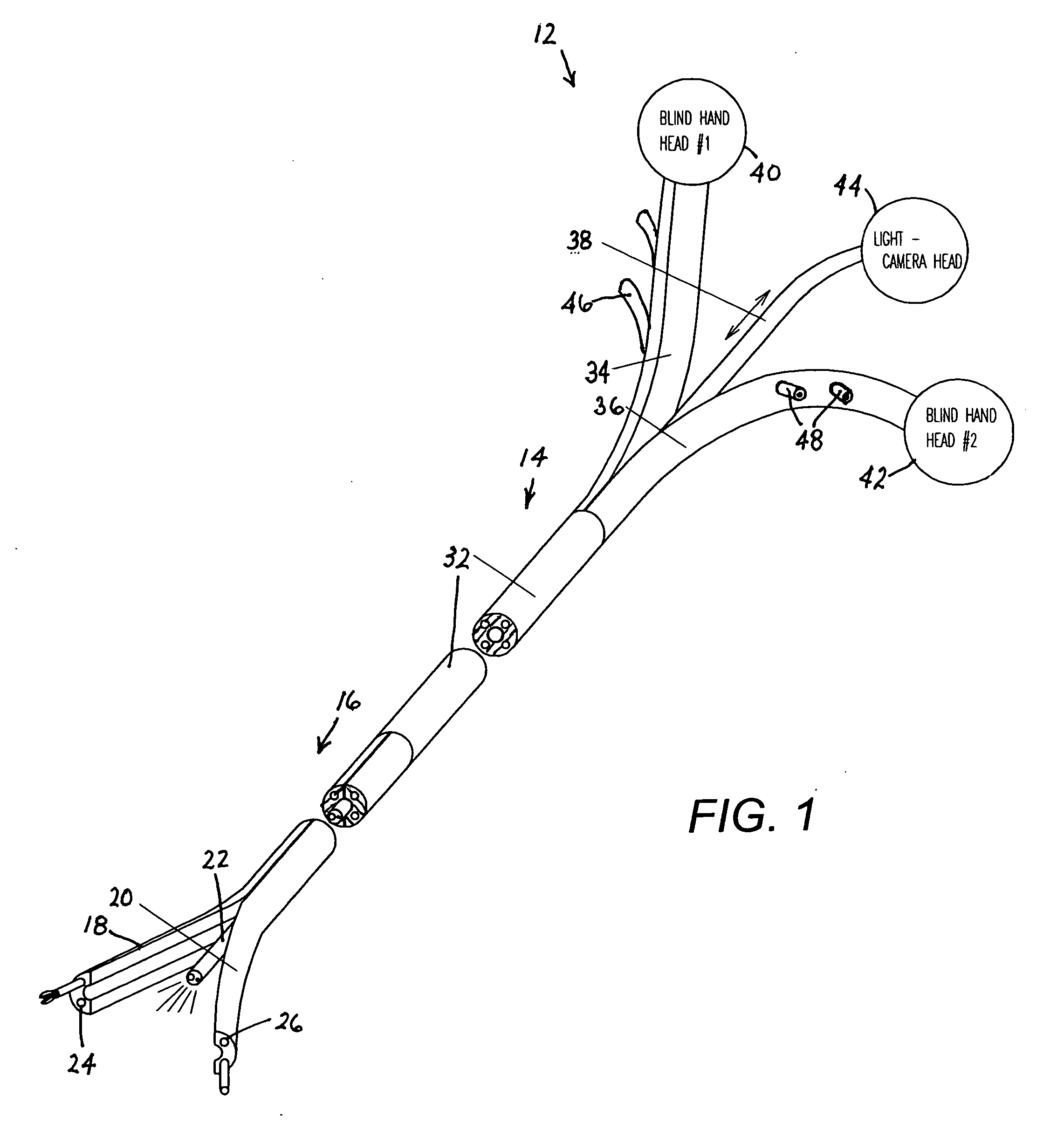
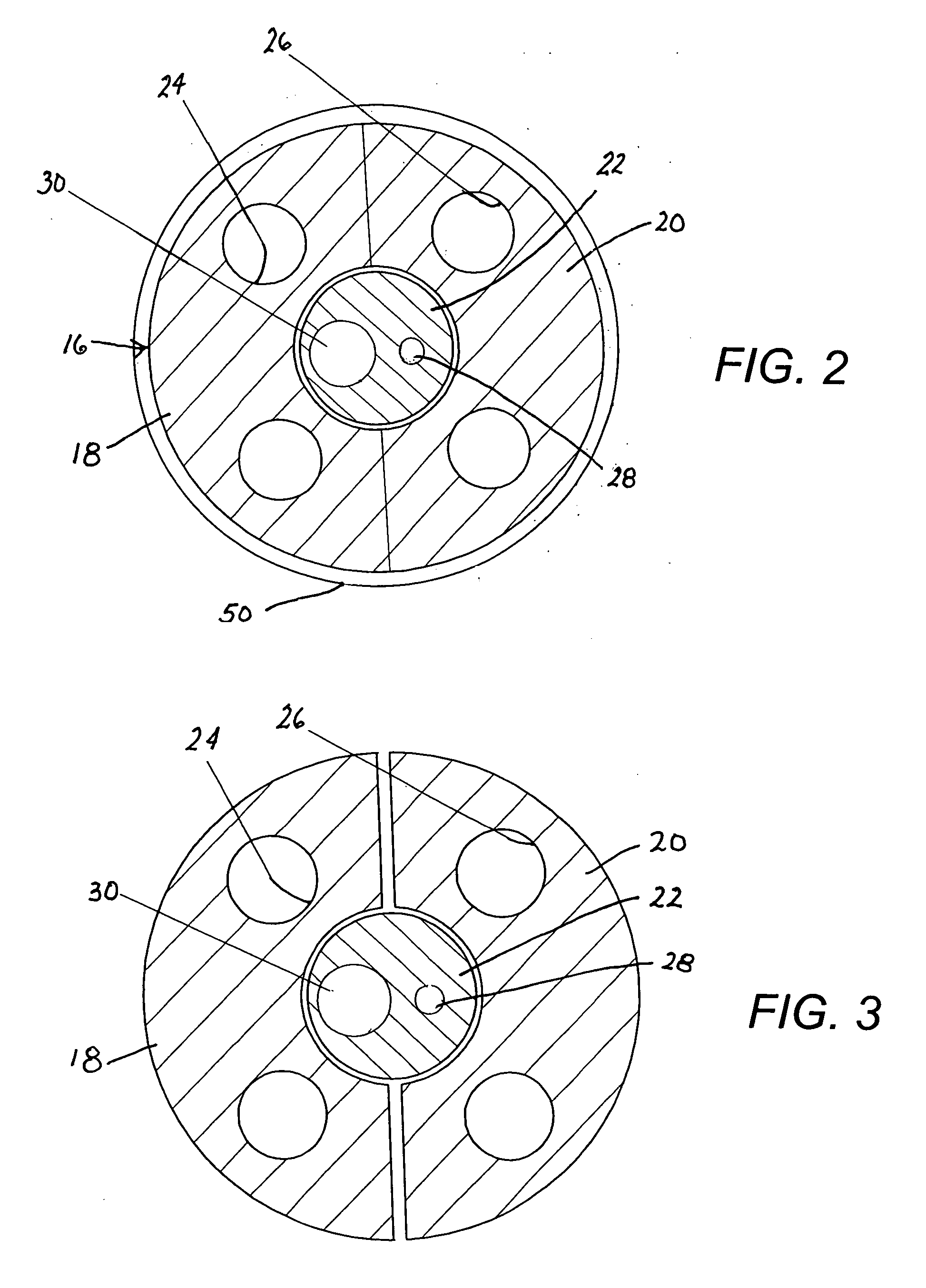
[0024] As depicted in FIG. 1, an endoscope 12 that is particularly useful for the performance of endoscopic surgical operations includes an insertion member 14 having a distal end portion 16 longitudinally split into a pair of working segments 18 and 20 and a central visualization segment 22. As shown in FIGS. 2 and 3, working segments 18 and 20 are each provided with a pair of longitudinal biopsy or working channels 24 and 26, whereas visualization segment 22 is provided with an illumination guide 28 and a lens 30 forming a portion of image transmission optics. The image transmission optics generally include a solid-state camera (not shown) generating an electrical image-encoding signal transmitted to a video monitor (not shown) for enabling visualization of a surgical site by operating surgeons or endoscopists. Visualization segment 22 may be additionally provided with a longitudinal channel (not illustrated) for conducting an irrigation liquid such as saline solution

[0025] As fu...


PUM


Abstract

A flexible fiberoptic endoscope has an insertion member with a distal end portion split longitudinally into a plurality of independently operable working segments each provided with at least one longitudinally extending working channel. Visualization optics are provided in the working segments or a separate central segment surrounded by the working segments. A sheath temporarily joins the working segments to one another during an introduction procedure. The sheath is removed upon insertion of the endoscope, so that the working segments may be / separated from one another and independently maneuvered via proximal control heads to facilitate the performance of an endoscopic surgical procedure.

Description

BACKGROUND OF THE INVENTION [0001] This invention relates to flexible fiberoptic endoscopy. More particularly, this invention relates to a fiberoptic endoscope and a related endoscopy technique. [0002] Flexible fiberoptic endoscopy is a minimally invasive procedure that is diagnostic for the most part. Certain minimal surgical procedures are being performed, but these are quite limited in scope. These surgical procedures include biopsy, polypectomy, and endoscopic mucosal resection (a recently introduced procedure). There are certain instruments used to endoscopically treat gastroesophageal reflux disorder (GERD. These instruments are large and clumsy and are, for the most part, attached to the endoscope rather than manipulated through its working channels. [0003] The main reason for the inability to perform surgical procedures through a flexible fiberoptic endoscope is that the endoscope itself represents “one hand.” One cannot suture (push and pull a needle), tie a knot, handle a ...

Claims


Application Information

Patent Timeline
Patent Type & Authority Applications(United States)
IPC IPC(8): A61B1/00A61B1/018A61B1/04A61B1/273A61B17/04A61B17/34
CPCA61B1/00071A61B1/0008A61B1/00105A61B1/00135A61B1/00142A61B1/0125A61B2017/3445A61B1/042A61B1/2736A61B17/0467A61B17/0469A61B2017/0475A61B1/018
Inventor NAKAO, NAOMI L.
Owner GRANIT MEDICAL INNOVATION
Learn More