
Computer readable recording medium recorded with diagnosis supporting program, diagnosis supporting apparatus and diagnosis supporting method

a computer-readable recording medium and support program technology, applied in diagnostic recording/measuring, instruments, applications, etc., can solve the problems of many problems still left, image diagnoses based on only subjective estimations fail to avoid misdiagnosis, and it is extremely difficult to select suitable reference images out of a great deal of stored ct images

Inactive Publication Date: 2002-05-30
FUJITSU LTD
View PDF3 Cites 58 Cited by
  • Summary
  • Abstract
  • Description
  • Claims
  • Application Information

AI Technical Summary

Benefits of technology

[0007] According to such a constitution, reference images which are image-wise similar to the diagnosis target image are retrieved from those reference images stored in the database, based on the feature quantities of the lesion position detected from the diagnosis target image. Thus, a doctor to conduct diagnosis by interpreting the diagnosis target image is possible to readily refer to those past disease cases similar to the disease case of the lesion portion appearing in the diagnosis target image. This allows objective diagnosis excluding the subjectivity. Since the objective diagnosis becomes possible, there can be drastically reduced the possibility of misdiagnosis even by a less experienced doctor, for example, to thereby enable an improvement of diagnosis precision. Further, even when a disease name of the diagnosis target image is unclear from the diagnosis target image only, the disease name can be estimated by utilization of reference disease cases, to thereby improve diagnosis precision.
[0010] According to such a constitution, similarities between the diagnosis target image and each of the reference images are defined based on the image-wise similarities, respectively, so that reference images are sequentially retrieved from those having higher similarities. Thus, those reference images having higher possibilities of usefulness are referred to when interpreting the diagnosis target image, to thereby avoid useless reference and to thereby improve the diagnosis efficiency. Here, if the similarities are calculated taking account of the weighting set for each organ, similarities corresponding to the characteristic of each organ are calculated, to thereby improve calculation precision of similarities. If the variably constituted table is set with the weighting, it becomes possible to conduct, for example, a correction corresponding to peculiar characteristics of a CT apparatus, thereby further improving the calculation precision of similarities.
[0011] Further, it is preferable to display the findings related to the retrieved reference images. In this way, the findings of the reference images can be referred to in addition to the reference images themselves, so that, even when a disease name for a patient is unclear, such a disease name can be diagnosed from the findings of similar disease cases.
[0013] According to such a constitution, the detecting procedures for organs which are not diagnosis targets are avoided, to thereby improve the processing speed. Further, by detecting, as the image-wise feature quantities, the global feature quantity, the topical feature quantity and the common feature quantity, it becomes possible to extract feature quantities corresponding to characteristics of each disease, thereby preventing oversight of a tumor, for example.

Problems solved by technology

However, image diagnoses based on only subjective estimations fail to avoid misdiagnoses such as due to oversight and / or misunderstanding.
However, many problems are still left such as due to the restriction of time.
However, it has been extremely difficult to select suitable reference images out of a great deal of stored CT images, since the conventional PACS merely allows to accumulate and refer to digital images.
As such, irrespective of the development of the PACS, since those CT images photographed in the past have not been fully utilized, diagnoses based on subjective estimations by doctors have been still conducted, resulting in a situation difficult for an improvement of diagnosis precision.

Method used


Image

  • Computer readable recording medium recorded with diagnosis supporting program, diagnosis supporting apparatus and diagnosis supporting method
  • Computer readable recording medium recorded with diagnosis supporting program, diagnosis supporting apparatus and diagnosis supporting method
  • Computer readable recording medium recorded with diagnosis supporting program, diagnosis supporting apparatus and diagnosis supporting method

Examples


Embodiment Construction

[0026] The present invention will be described hereinafter with reference to the accompanying drawings.

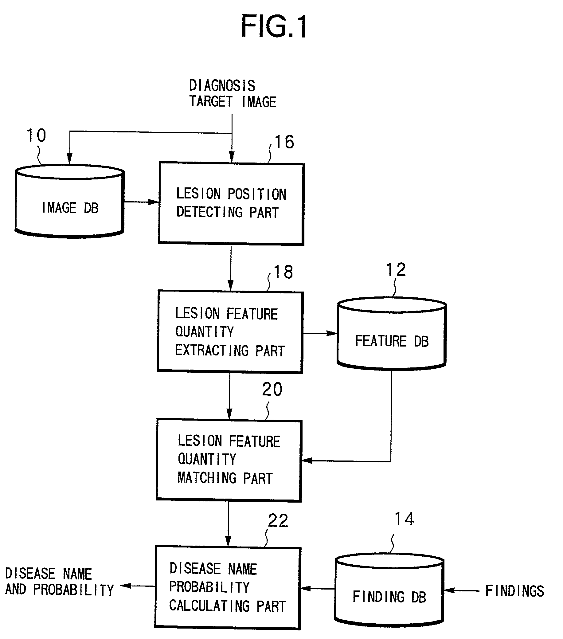
[0027] FIG. 1 shows a constitution of a diagnosis supporting apparatus realizing the diagnosis supporting technique according to the present invention. The diagnosis supporting apparatus is constructed on a computer system comprising at least a central processing unit (CPU) and a memory, and operates according to a program loaded onto the memory.

[0028] The diagnosis supporting apparatus comprises an image database 10, a feature database 12, a finding database 14, a lesion position detecting part 16, a lesion feature quantity extracting part 18, a lesion feature quantity matching part 20 and a disease name probability calculating part 22. Note, the term "database" shall be abbreviated to "DB" in the following description.

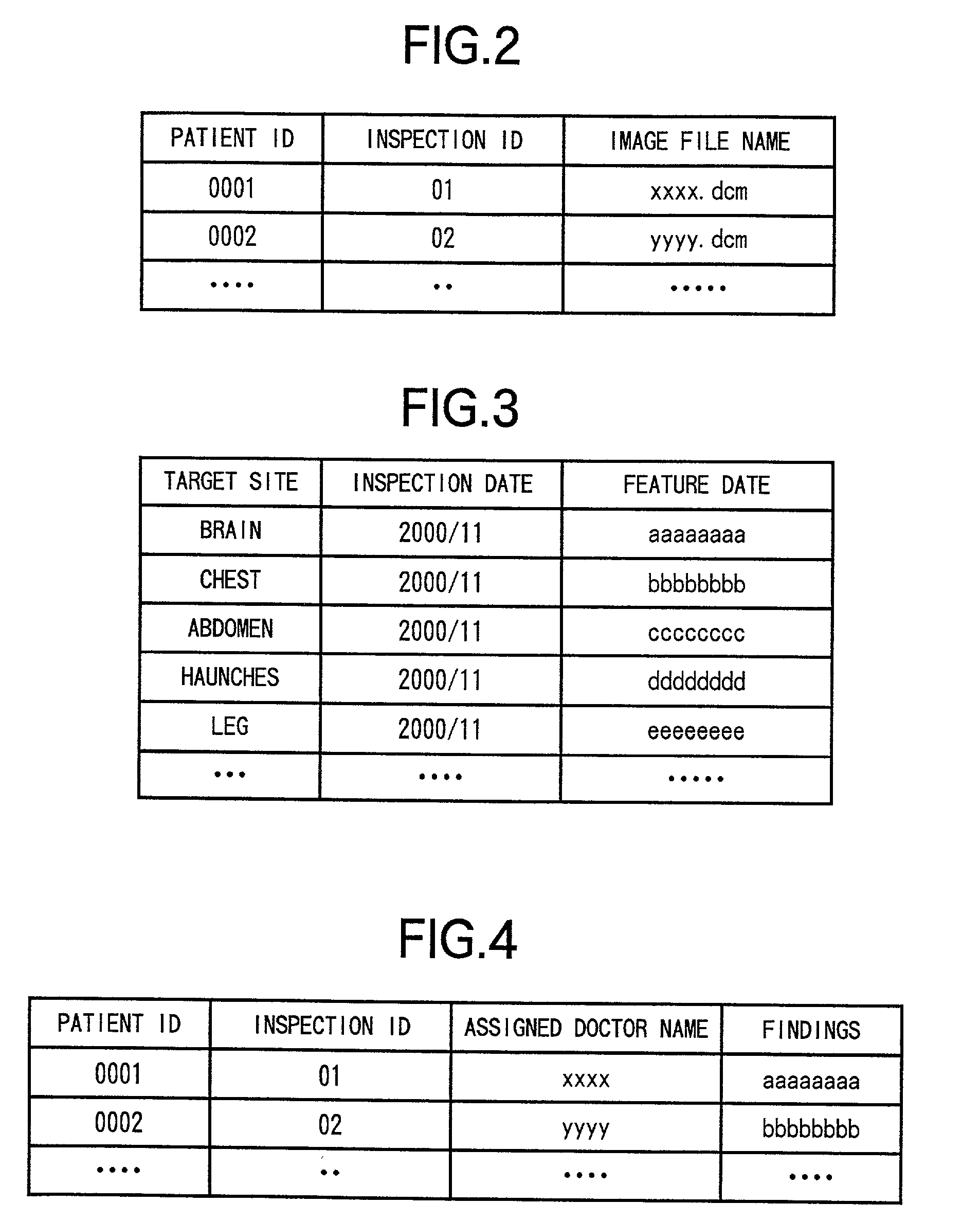
[0029] Stored in the image DB 10 are image data such as CT images and MRI images in the format in accordance with DICOM (Digital Imaging and Communications in Medic...


PUM


Abstract

A computer readable recording medium recorded with a diagnosis supporting program, a diagnosis supporting apparatus and a diagnosis supporting method, enabling to retrieve similar disease cases making use of feature quantities of CT images and / or MRI images when interpreting such images, to thereby allow an improvement of diagnosis precision.

Description

[0001] The present invention relates to a technique for improving diagnosis precision particularly in a diagnosis supporting technique for supporting diagnoses by image interpretation.RELATED ART OF THE INVENTION[0002] There have been conventionally conducted interpretation of CT (Computerized Tomography) images and MRI (Magnetic Resonance Imaging) images, for example, by subjective estimations based on experiences stored over time by doctors such as radiologists. However, image diagnoses based on only subjective estimations fail to avoid misdiagnoses such as due to oversight and / or misunderstanding. To avoid such misdiagnoses, various endeavors have been made such as by interpreting CT images by a plurality of doctors. However, many problems are still left such as due to the restriction of time.[0003] Meanwhile, digitization of CT images has been intensely developed at present, such that almost all CT images are digitized in the fields of from simple photographs to angiographic ima...

Claims


Application Information

Patent Timeline
IPC IPC(8): G06F19/00
CPCG06F19/321Y10S707/99936G06F19/3487G06F19/345G16H50/20G16H15/00G16H70/60G16H30/20
Inventor MURAO, KOHEI
Owner FUJITSU LTD
Learn More