
System and methods for fully automated data analysis, reporting and quantification for medical and general diagnosis, and for edge detection in digitized images

a fully automated, image-based technology, applied in image data processing, instruments, computing, etc., can solve the problems of large limitations in the full automation of image analysis, near-infinitely complex machines and machine operations, and accurate fully automated systems, so as to improve detection and increase or decrease the gain

Active Publication Date: 2021-04-13
EYES LTD
View PDF16 Cites 6 Cited by
  • Summary
  • Abstract
  • Description
  • Claims
  • Application Information

AI Technical Summary

Benefits of technology

[0027]Another object of the invention is to give the image capturing device or input module (such as camera or echo machine) the ability to automatically adapt based on the information given it to increase or decrease the gain in the area where the edge is deficient or excess, respectively to try to improve the detection.

Problems solved by technology

First, the use of functional / operational technical descriptions allows near-infinitely complex machines and machine operations arising from interchained hardware elements to be described in a manner that the human mind can process (e.g., by mimicking natural language and logical narrative flow).
Due to the huge amount of information presented in an image and the lack of accurate fully automated systems for analysis, the field of medicine is frequently left to semi-quantitation (e.g., grade on a 1 to 4 scale).
While this may be adequate for many purposes, observer bias, lack of reproducibility, and lack of the ability to be precise often results in the need for more testing to try to be certain of the diagnosis, or painstaking effort by the physician to make sure the treatment is given in the effective regions (e.g., that the proper location on the field of image is treated by X-rays, etc.).
Currently there have been great limitations in the full automation of image analysis.
None of the current technologies and prior art, taken alone or in combination, address nor provide a utility solution for fully automated diagnostic data analysis and image quality assessment based on the extraction of predefined parameters from the input of data to be used for further reporting as accurate quantitative measurements and qualitative inferences regarding medical and general diagnosis of patients.
The raw images obtained in these arts including cineangiograms of the coronary arteries and echo images of the cardiac chamber walls until presently have not readily been amenable to accurate fully automated analysis of artery caliber (diameter along its length to evaluate amount of stenosis) or chamber dimensions and wall thickness.
However, actual automation of this analysis is burdensome and currently has been inaccurate and therefore unreliable.
In echocardiography field, there is currently no fully automated method of measuring cardiac chamber borders and dimensions.

Method used


Image

  • System and methods for fully automated data analysis, reporting and quantification for medical and general diagnosis, and for edge detection in digitized images

Examples


Embodiment Construction

[0028]The novel features believed to be characteristics of the invention are set forth in the appended claims. The invention itself, however, as well as the preferred mode of use, further objects and advantages thereof, will best be understood by reference to the following detailed description of illustrative embodiment when read in conjunction with the accompanying drawings, wherein:

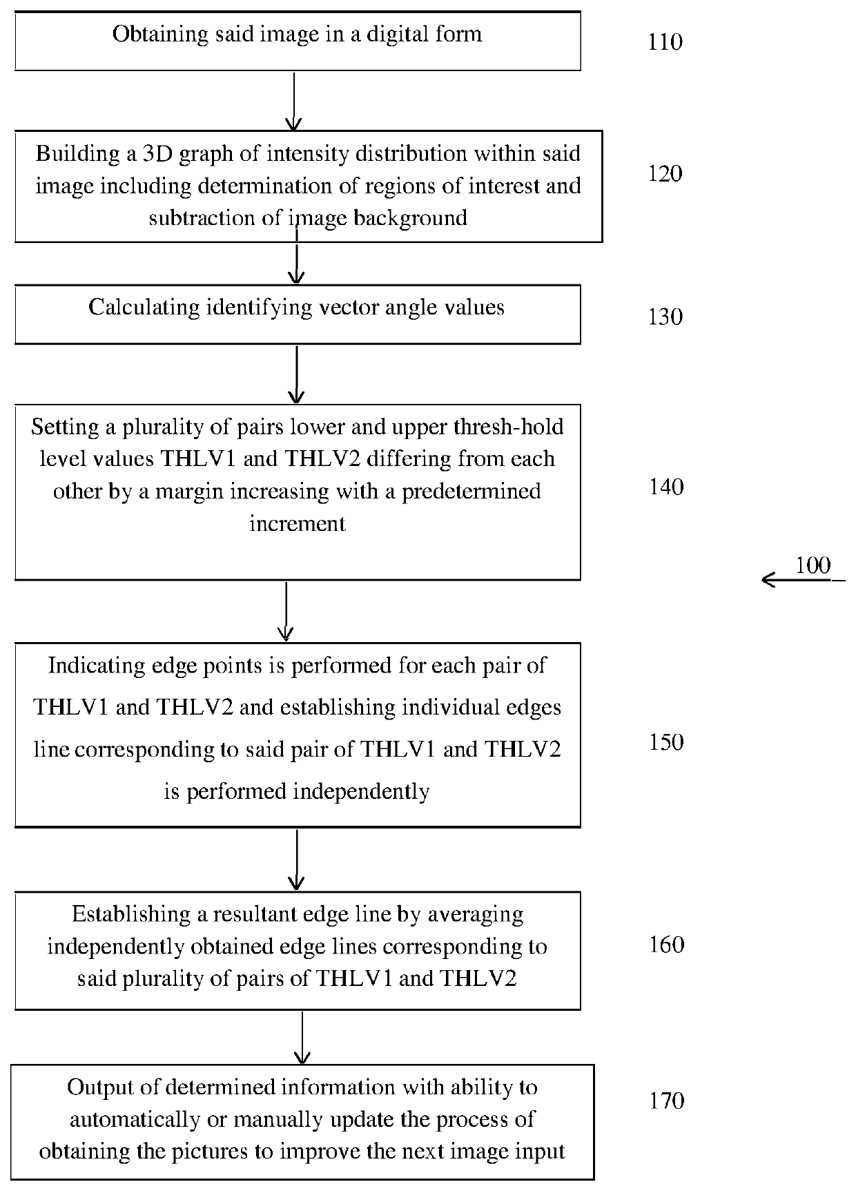
[0029]FIG. 1 is a flowchart of a method of detecting of an edge in digitized images

DETAILED DESCRIPTION OF THE PREFERRED EMBODIMENTS

[0030]In the following detailed description of the preferred embodiments, reference is made to the accompanying drawings that form a part hereof, and in which are shown by way of illustration specific embodiments in which the invention may be practiced. It is understood that other embodiments may be utilized and structural changes may be made without departing from the scope of the present invention. The present invention may be practiced according to the claims without som...


PUM


Abstract

A method of detecting an edge within digitized images, said method comprising steps of: (a) obtaining said image in a digital form; (b) building a 3D graph of intensity distribution within said image; (c) finding Regions of Interest ROI in the image and subtracting out the background; (d) establishing edge lines within said image; establishing a resultant edge line for each ROI by averaging or maximizing the set of independently obtained edge lines corresponding to said plurality of criteria but that differ from each other by a margin increasing with a predetermined increment; and (f) generating at least one report presenting the quantitative and / or qualitative parameters in a form of accurate quantitative measurements and qualitative inferences regarding medical and general diagnosis of the images and patients utilizing one or more output modules.

Description

RELATED APPLICATIONS[0001]This application is a National Phase of PCT Patent Application No. PCT / IL2017 / 051344 having International filing date of Dec. 13, 2017, which claims the benefit of priority of U.S. Provisional Application No. 62 / 433,815 filed on Dec. 14, 2016. The contents of the above applications are all incorporated by reference as if fully set forth herein in their entirety.FIELD OF THE INVENTION[0002]The present invention relates generally to the field of image assessment and analysis. More specifically, the present invention relates to edge detecting in digitized images.BACKGROUND OF THE INVENTION[0003]The following description includes information that may be useful in understanding the present invention. It is not an admission that any of the information provided herein is prior art or relevant to the presently claimed invention, or that any publication specifically or implicitly referenced is prior art.[0004]Importantly, although the operational / functional descript...

Claims


Application Information

Patent Timeline
Patent Type & Authority Patents(United States)
IPC IPC(8): G06T7/13G06T7/162G06T7/194
CPCG06T7/13G06T7/162G06T7/194G06T2207/20032G06T2207/20072G06T2207/30048G06T2207/30101
Inventor CHADI, BENJAMIN H.
Owner EYES LTD
Learn More