Method for the rapid and convenient detection and enumeration of neutrophils in biological samples

a biological sample and neutrophil technology, applied in the field of inflammatory conditions, can solve the problems of general limitation of methods for identification and enumeration of neutrophils, lack of specific antigenic cross-species markers for this cell type,

Active Publication Date: 2020-07-21
MEP EQUINE SOLUTIONS
View PDF2 Cites 0 Cited by
  • Summary
  • Abstract
  • Description
  • Claims
  • Application Information

AI Technical Summary

Benefits of technology

[0024]The purpose of this invention is to provide a rapid (under 10 minute) method to image large numbers of cells (containing neutrophils) and then automatically count the neutrophils without the need for a trained operator or bulky / delicate / expensive equipment.
[0026]The core of this invention is a method of specifically rendering neutrophils fluorescent and coupling this method to a device capable imaging the cells and counting either the total neutrophils or both the total number of neutrophils and somatic cells in milk or other bodily fluids. Dual-wavelength fluorescence imaging of neutrophils and (optionally) nuclei facilitates DSCC (and optionally also SCC) in a single device and so provides enhanced sensitivity for early infection detection over SCC alone. This invention can also be incorporated into a four-quarter test of udder milk in ruminant animals.
[0029]However, color changes in the visible range provide insufficient contrast to identify specific cells unless viewed under high magnification, which would be incompatible with a portable device and would also require a substantially more sophisticated computer vision algorithm in order to accurately identify neutrophils. This invention utilizes this same enzyme to generate a fluorescent, insoluble product within the neutrophils themselves. As a result, the high-contrast of fluorescence-mode detection means cells can be imaged at much lower magnification (and even de-magnification). This not only reduces the complexity and fragility of the optical system required for imaging, but also reduces the size and weight of the device. Furthermore, by imaging at lower magnification increases the field-of-view and allows for the instantaneous imaging of more cells, thereby increasing the accuracy / precision by increasing the number of countable cells. Finally, and unlike chromogenic histological tests, because the substrates are non-fluorescent while the product is, no separation of staining solution from sample is required prior to imaging.
[0032]However, this staining still requires a bulky high-magnification microscope to manually identify and enumerate neutrophils, which is tedious, very time consuming and requires specialized training. Automation of counting would require a motorized stage to take multiple photographs for analysis, due to the limited field of view, adding to the cost and complexity of the device, reducing its portability and increasing the assay time due to such data collection restrictions.
[0033]Imaging more cells in a single field requires lower power optics (perhaps even less than 1×), which in turn results in a smaller and both a more economical and portable unit. Additional field-of-view and resolving power can be provided, if required, by increasing the resolution of the image sensor without compromising portability. Unfortunately, visible light microscopy is not conducive to reducing magnification indefinitely without significant loss in fidelity because of its limited signal-to-noise ratio. On the other hand, fluorescence microscopy affords the advantage of excellent signal-noise. In fact, several cell-counting devices exist that label cellular nuclei with fluorescent DNA intercalating reagents and produce images for automated enumeration where the “cells” appear as objects only 1-5 pixels in size (e.g., Nucleocounter, Chemometec and Quick SCC, Dairy Quality Inc.; see above and FIG. 2).
[0040]It should be noted that other systems have been described for the fluorogenic detection of cellular esterase activity, but that these have in general been limited to the detection viable cells or multiple cell populations as opposed to specific cell types66. In contrast, this invention focuses on the utilization of a specific esterase activity that is present in a specific subset of clinically relevant cells. More specifically, this invention focuses on CAE, which to date has only been detected optically using standard histological methods. This invention improves on the current art but introducing a high-contrast, low signal-noise fluorescence detection modality for CAE and coupling it to image capture at low magnification to maximize field-of-view, and then to computation image analysis in order to automate and accelerate the acquisition of quantitative data.

Problems solved by technology

One difficulty in developing a device to detect neutrophils is the dearth of specifics antigenic cross-species markers for this cell type.
Without a specific antigen to provide a means to differentiate neutrophils and other cell types, their identification and enumeration are generally limited to the methods described above.

Method used


Image

  • Method for the rapid and convenient detection and enumeration of neutrophils in biological samples
  • Method for the rapid and convenient detection and enumeration of neutrophils in biological samples
  • Method for the rapid and convenient detection and enumeration of neutrophils in biological samples

Examples


Embodiment Construction

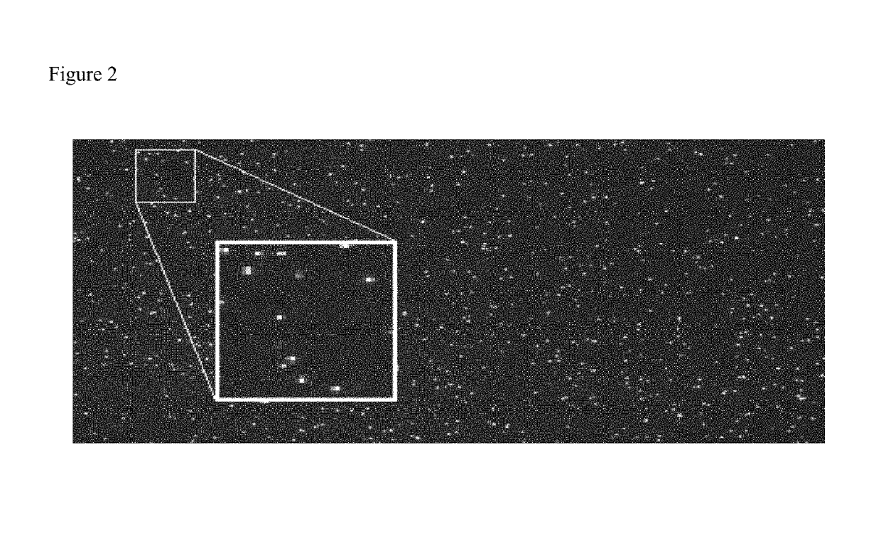
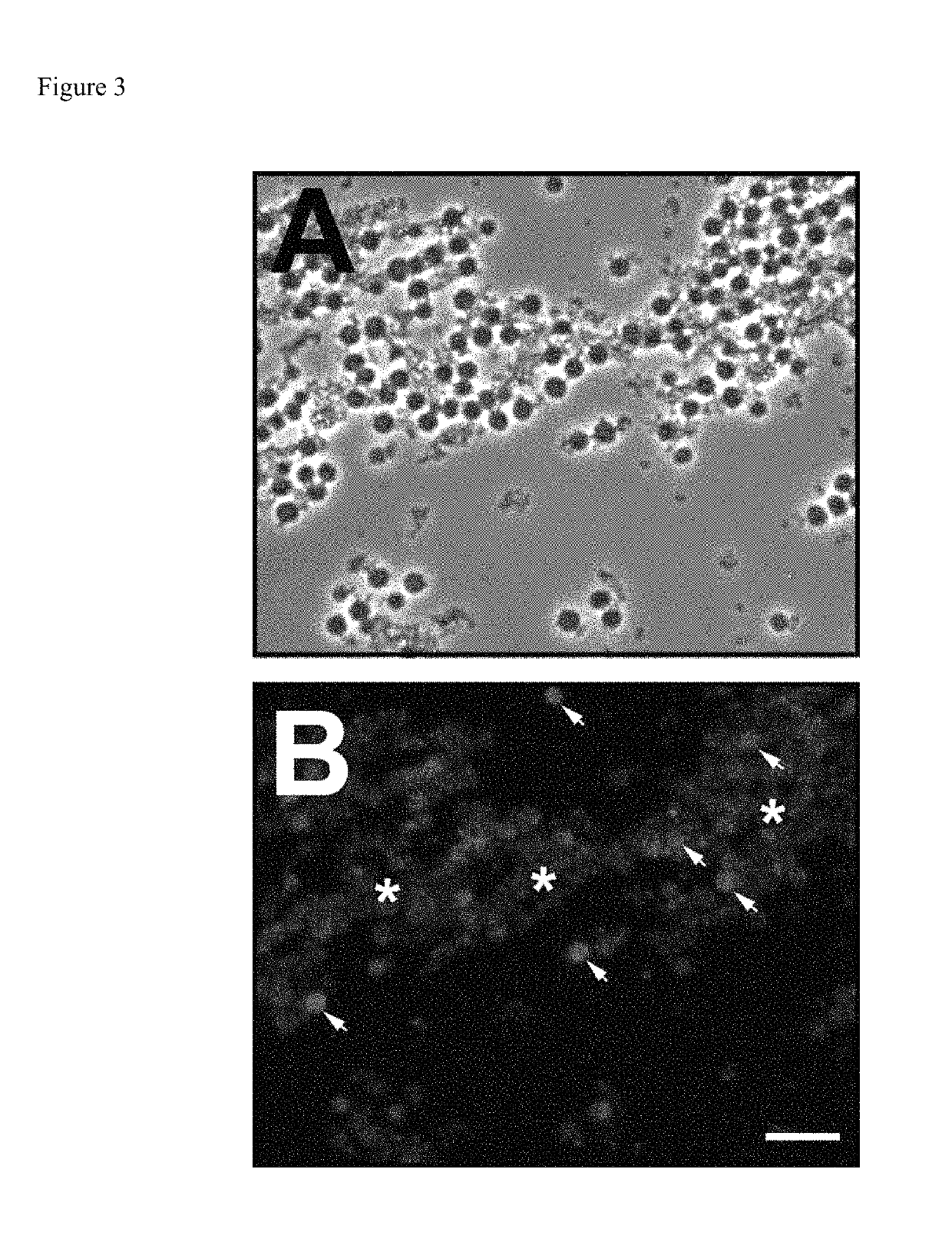
[0053]CAE activity is commonly detected using the Fast Red (FR) series of diazonium salts, though other salts, such as nitrosylated pararosanilin and Fast Corinth V, are used to produce azo dyes of different colors. Furthermore, not all diazonium salts produce azo dyes with the same (or any) fluorescent properties63. Any diazonium salt capable of reacting the CAE cleavage product of NCA to generate a fluorescent moiety could be used for the purposes of practicing this invention. Because FRTR has, however, been shown to produce an intensely fluorescent product following reaction a dephosphorylated naphthol phosphate in a phosphatase assay, upon conceiving of the invention, the inventor tested the ability of CAE to render neutrophil cells isolated from equine peripheral blood fluorescent after treatment with a combination with NCA (0.2 mg / ml) and FRTR (2 mg / ml) in a buffer of phosphate buffered saline (PBS).

[0054]While this mixture did indeed produce fluorescent material when examined...


PUM

PropertyMeasurementUnit
diameteraaaaaaaaaa
concentrationaaaaaaaaaa

Abstract

Methods are provided to facilitate the detection and enumeration of neutrophils in bodily fluids, including milk. The methods incorporate the fluorescent staining of neutrophils using fluorogenic enzyme substrates, the imaging of fluorescent cells using a digital device, and the electronic counting of the cells.

Description

CLAIM OF PRIORITY[0001]This application claims the benefit of U.S. Provisional Patent Application No. 62 / 484,014 filed Apr. 11, 2017.FIELD OF THE INVENTION[0002]This document relates to the diagnosis of inflammatory conditions and describes methods for quantifying the number of neutrophils in body fluids, including milk.BACKGROUND[0003]The process of inflammation is characterized by the rapid influx of a particular granulocyte, the neutrophil, into the affected area as a response to either physical injury or pathogen invasion1,2. Detection of elevated neutrophil levels, therefore, represents a marker for numerous inflammatory events and conditions that can be useful in the diagnosis and subsequent treatment of disease. Three examples of areas in which detection of neutrophils is of clinical value are intra-mammary mastitis in cows and other domesticated herbivores3, in acute respiratory distress syndrome (ARDS) in humans and in equine inflammatory airway disease (EIAD)4.[0004]Tradit...

Claims


Application Information

Patent Timeline
Patent Type & Authority Patents(United States)
IPC IPC(8): G01N33/50G01N15/10G01N33/04G01N31/22G01N15/06G01N21/64G01N15/14
CPCG01N15/06G01N33/5094G01N21/6428G01N15/1463G01N33/04G01N2015/1006G01N2015/0693G01N31/22G01N2015/1486G01N2021/6439
Inventor SLUSAREWICZ, PAWEL
Owner MEP EQUINE SOLUTIONS
Why Eureka
Try Eureka