Identifying abnormal tissue in images of computed tomography

a computed tomography and abnormal tissue technology, applied in image enhancement, instruments, applications, etc., can solve the problem of low contrast between abnormal tissue and blood vessels containing contrast medium, and achieve the effect of simplifying the subsequent analysis

Active Publication Date: 2019-10-01
KONINKLJIJKE PHILIPS NV
View PDF39 Cites 0 Cited by
  • Summary
  • Abstract
  • Description
  • Claims
  • Application Information

AI Technical Summary

Benefits of technology

[0006]In accordance with the invention, an imaging method for identifying abnormal tissue in the lung is provided, comprising the recording of slice images of the lung by means of X-ray radiation, recording of blood vessels, differentiation of blood vessels and abnormal tissue, segmentation of the abnormal tissue and display of the segmented abnormal tissue on an output device. Furthermore, a computer tomograph for identifying abnormal tissue in the lung is provided, having a radiation source for recording slice images of the lung and blood vessels by means of X-ray radiation, a computer unit for differentiating the blood vessels from abnormal tissue and for segmenting the abnormal tissue, as well as an output device for displaying the segmented abnormal tissue. In addition, a computer program for controlling a computer tomograph for identifying abnormal tissue in the lung and having a radiation source for recording slice images of the lung and of blood vessels by means of X-ray radiation is provided, for differentiating the blood vessels from abnormal tissue, for segmenting the abnormal tissue and for controlling an output device for displaying the abnormal tissue. By means of the invention, abnormal tissue in the vicinity of blood vessels is distinguishable from these. This appreciably simplifies the subsequent analysis of the results of the imaging method.

Problems solved by technology

The problem with this process, however, is that the contrast between abnormal tissue and the blood vessels containing the contrast medium is low, the attenuation values, also known as Hounsfield values, received by the detector device of the computer tomograph do not differ significantly from one another.

Method used


Image

  • Identifying abnormal tissue in images of computed tomography
  • Identifying abnormal tissue in images of computed tomography
  • Identifying abnormal tissue in images of computed tomography

Examples


Embodiment Construction

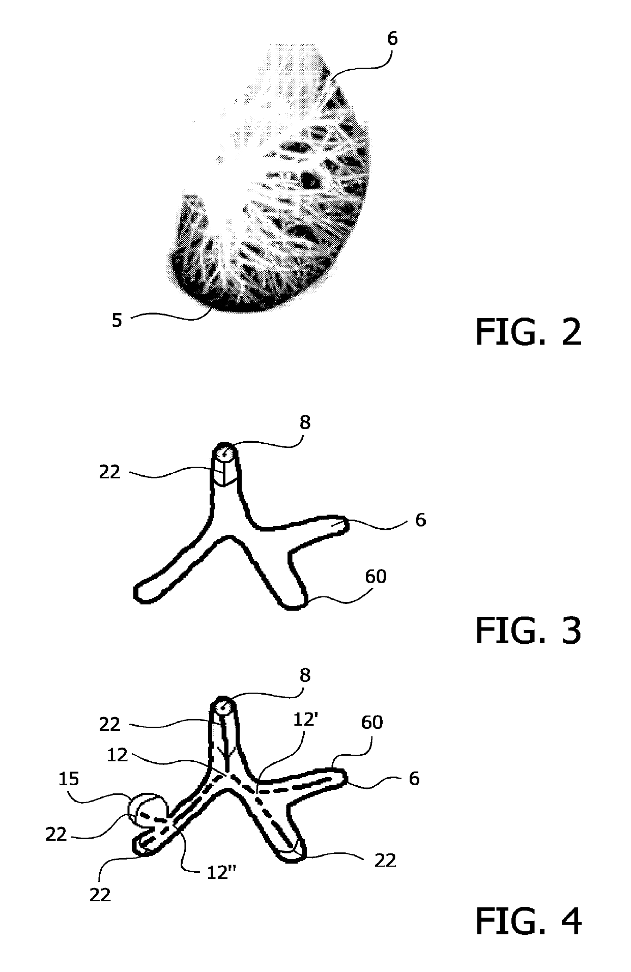
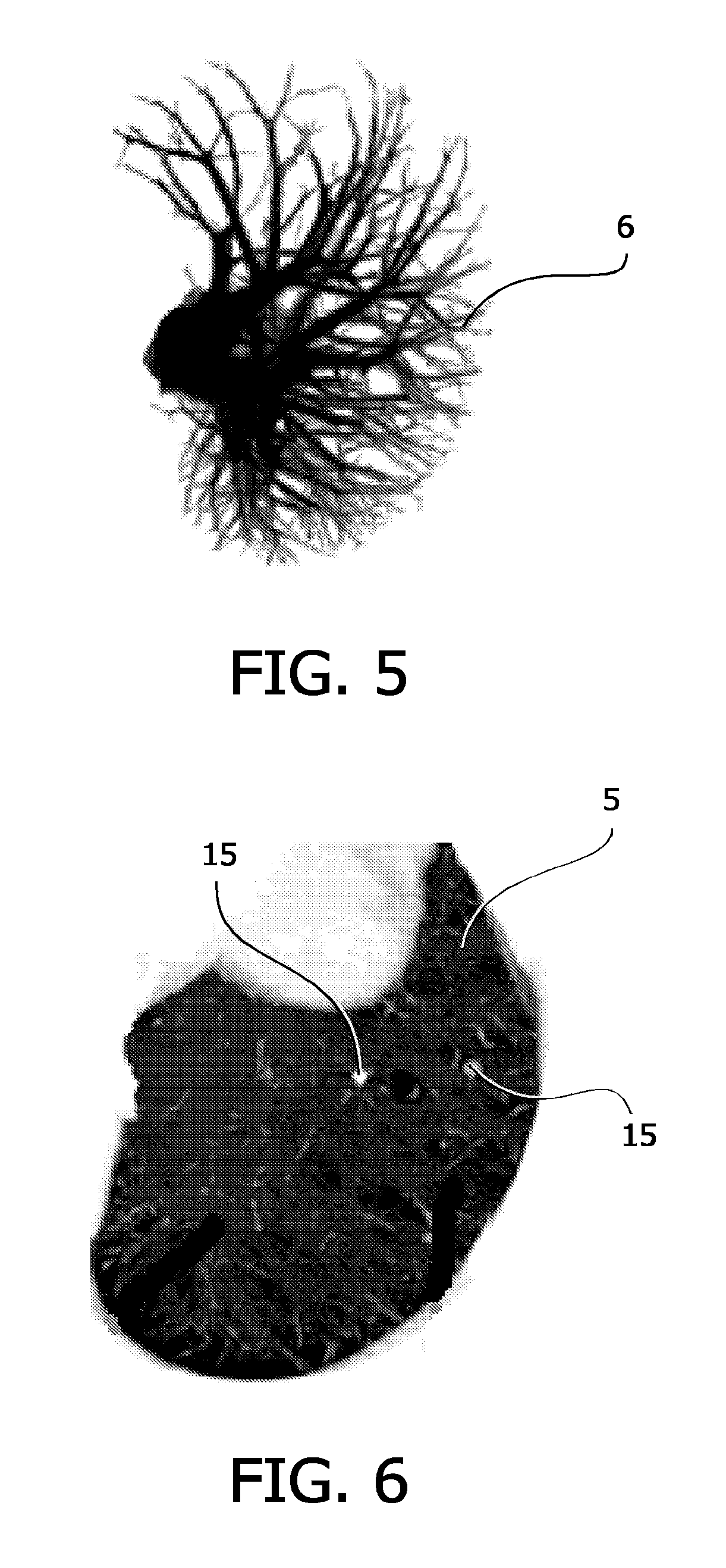
[0021]FIG. 1 shows a schematic view of a support or a gantry 1, which is able to rotate along the circular path marked by the curved arrow parallel to the y-plane of the coordinate system illustrated. For that purpose, the gantry 1 is driven by a motor 2 at a preferably constant but adjustable angular velocity. A radiation source 20, for example an X-ray tube, is fixed to the gantry 1, and includes a collimator arrangement, which extracts a fan-shaped beam bundle 4 from the radiation produced by the radiation source 20. The beam bundle 4 is illustrated schematically by means of two lines, which bound the beam bundle 4. The fan-shaped beam bundle 4 penetrates at least partially through an object 13, which in FIG. 1 is shown as a portion of a cylinder; the object 13 is usually a patient or part of a patient on a patient support table and in this case the object comprises a lung or portions thereof, the lung cavity 5, which comprises the lung, pulmonary tissue and blood vessels 6, whic...


PUM


Abstract

An imaging method for identifying abnormal tissue in the lung is provided, comprising the recording of slice images of the lung by means of X-ray radiation, recording of blood vessels, differentiation of blood vessels and abnormal tissue, segmentation of the abnormal tissue and display of the segmented abnormal tissue on an output device. In addition, a computer tomograph for identifying abnormal tissue in the lung is provided, having a radiation source for recording slice images of the lung and blood vessels by means of X-ray radiation, a computer unit for differentiating the blood vessels from the abnormal tissue and for segmenting the abnormal tissue, as well as an output device for displaying the segmented abnormal tissue. Furthermore, a computer program is provided for controlling a computer tomograph for an identification of abnormal tissue in the lung by means of a radiation source, designed to record slice images of the lung and blood vessels by means of X-ray radiation, to differentiate the blood vessels from abnormal tissue, to segment the abnormal tissue and to control an output device for displaying the abnormal tissue.

Description

CROSS REFERENCE TO RELATED APPLICATIONS[0001]This application is a divisional of U.S. application Ser. No. 11 / 815,973 filed Aug. 10, 2007, which is a national filing of international application serial number PCT / IB2006 / 050362, filed Feb. 3, 2006, which claims the benefit of EP application serial number 05101024.7 filed Feb. 11, 2005, all of which are incorporated herein by reference.FIELD OF THE INVENTION[0002]The invention relates to an imaging method for computed, to a computer tomograph and to a computer program.BACKGROUND OF THE INVENTION[0003]In the technical field of medical radiology, computer tomographs have gained increasingly in importance in recent years. In computed tomography, x-ray radiation is irradiated from a radiation source towards an object to be examined, as a rule a patient, and, on the basis of the attenuation of the X-ray radiation after this has passed through the object to be examined, an image is produced on a monitor. In this process, the radiation sourc...

Claims


Application Information

Patent Timeline
Patent Type & Authority Patents(United States)
IPC IPC(8): G06T7/00G06T7/10G06T5/50G06T7/149G06T7/12A61B6/00A61B6/03
CPCG06T7/12G06T7/10G06T7/149A61B6/5217G06T5/50G06T7/0012G06T2207/30101G06T2207/10116A61B6/032G06T2207/30064G06T2207/30061A61B6/481G06T2207/10081
Inventor BUELOW, THOMASWIEMKER, RAFAELLORENZ, CRISTIANRENISCH, STEFFENBLAFFERT, THOMAS
Owner KONINKLJIJKE PHILIPS NV
Why Eureka
Try Eureka