Devices useable for treatment of glaucoma and other surgical procedures

a technology for glaucoma and other surgical procedures, applied in the field of glaucoma, can solve the problems of abnormally high intraocular pressure, increased pressure of aqueous humor within the anterior chamber, and sometimes unsuccessful trabeculoplasty, trabeculectomy and shunt implantation procedures

Active Publication Date: 2018-11-13
MICROSURGICAL TECH INC
View PDF69 Cites 6 Cited by
  • Summary
  • Abstract
  • Description
  • Claims
  • Application Information

AI Technical Summary

Benefits of technology

The invention provides a device and method for cutting or ablating tissue in a patient's body. By controlling the amount of power or energy delivered to the tissue cutting or ablation apparatus, the device can control the size and extent of the high power density zone and the surrounding medium power density zone, resulting in cutting or ablation of a strip of tissue of a desired size while avoiding significant or irreparable damage to tissue that is to be protected or other tissue at the margins of or in close proximity to the strip of tissue that has been cut or ablated. The device can be used in percutaneous surgical procedures, such as goniectomy procedures, where it is inserted through the trabecular meshwork into Schlemm's Canal.

Problems solved by technology

Glaucoma is frequently, but not always, accompanied by abnormally high intraocular pressure.
However, in many glaucoma patients, drainage of the aqueous humor through the trabecular meshwork is impaired, thereby causing the pressure of aqueous humor within the anterior chamber to increase.
Trabeculoplasty, trabeculectomy and shunt implantation procedures are sometimes unsuccessful due to scarring of closure of the surgically created channels or holes and / or clogging of the drainage tube.

Method used


Image

  • Devices useable for treatment of glaucoma and other surgical procedures
  • Devices useable for treatment of glaucoma and other surgical procedures
  • Devices useable for treatment of glaucoma and other surgical procedures

Examples


Embodiment Construction

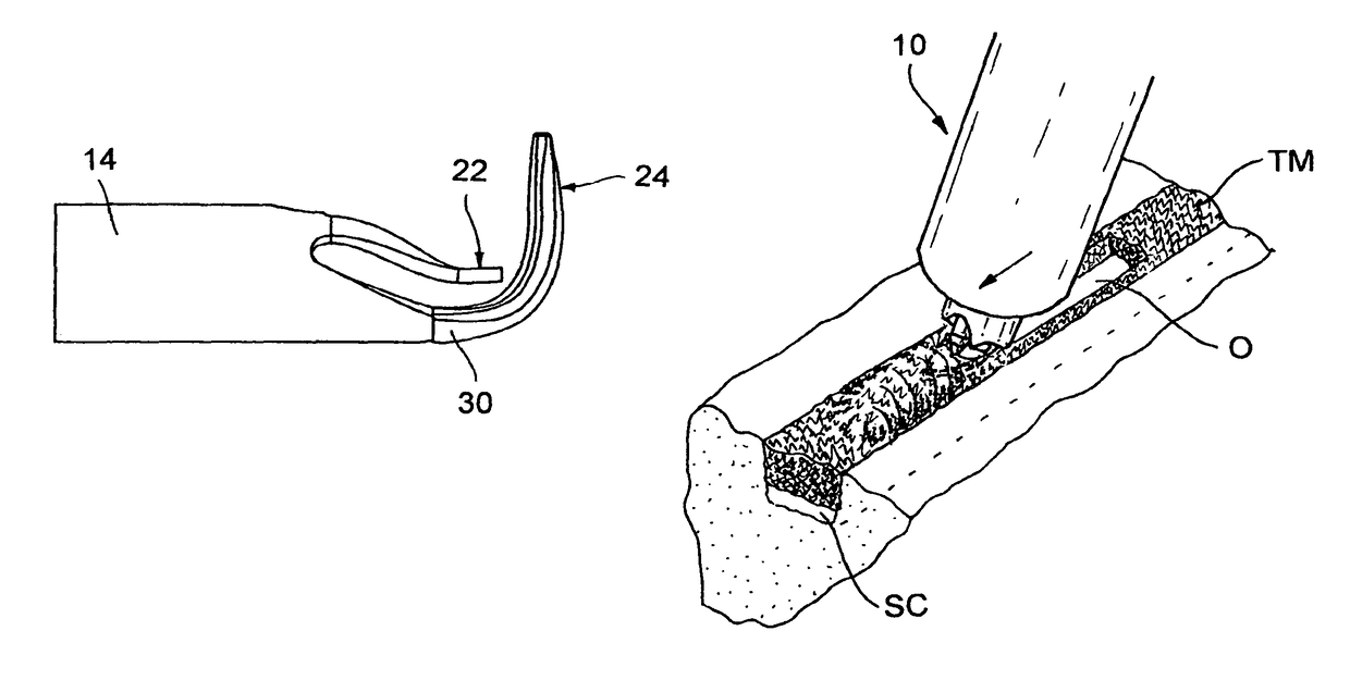
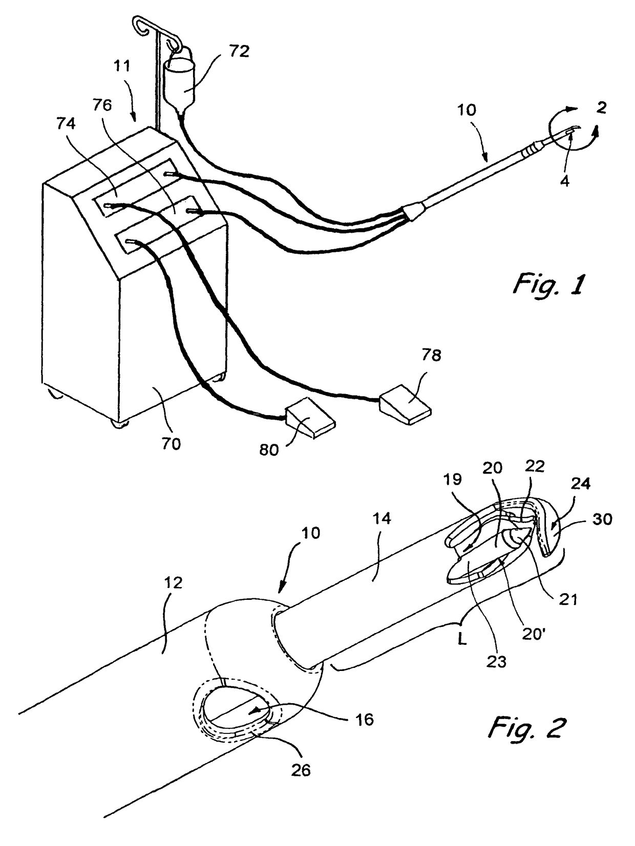
[0038]Turning now to FIG. 1, a device in accordance with the present invention for cutting and / or ablating tissue, for example, tissue of an eye during a goniectomy procedure, is shown generally at 10. The device 10 generally comprises an elongate handpiece or probe having a distal end having a tissue cutting or ablating apparatus 4 disposed generally within the distal end or distal portion of the probe 3. The tissue cutting or ablating apparatus 4 may be a suitable mechanism designed to cut, ablate, cauterize, sever and / or remove tissue from a target region, for example, from a surgical site. The device 10 may be part of a larger surgical system 11, for example, the device 10 may be structured and adapted to be operatively connectable to a separate apparatus, for example a surgical control console 70 for controlling and powering operation of various functions of the device during a surgical procedure. Examples of surgical consoles that may be suitable include but are not limited to...


PUM


Abstract

A device and method for cutting or ablating tissue in a human or veterinary patient includes an elongate probe having a distal end, a tissue cutting or ablating apparatus located adjacent within the distal end, and a tissue protector extending from the distal end. The protector generally has a first side and a second side and the tissue cutting or ablating apparatus is located adjacent to the first side thereof. The distal end is structured to be advanceable into tissue or otherwise placed and positioned within the patient's body such that tissue adjacent to the first side of the protector is cut away or ablated by the tissue cutting or ablation apparatus while tissue that is adjacent to the second side of the protector is not substantially damaged by the tissue cutting or ablating apparatus.

Description

RELATED APPLICATIONS[0001]This application is a division of copending U.S. patent application Ser. No. 10 / 560,266 filed May 11, 2006, which is a 35 U.S.C. § 371 national stage of PCT International Patent Application No. PCT / US2004 / 018483 Filed Jun. 10, 2004, which claims priority to U.S. Provisional Patent Application No. 60 / 477,258 filed Jun. 10, 2003, the entire disclosure of each such application being expressly incorporated herein by reference.BACKGROUND OF THE INVENTION[0002]A. Symptoms and Etiology of Glaucoma[0003]The term “glaucoma” refers generally to a group of diseases which cause progressive damage to the optic nerve and resultant optical field defects, vision loss and, in some cases, blindness. Glaucoma is frequently, but not always, accompanied by abnormally high intraocular pressure. Aqueous humor is continually produced by cells of the ciliary body and such aqueous humor fills the anterior chamber of the eye. Excess aqueous humor normally drains from the anterior cha...

Claims


Application Information

Patent Timeline
Patent Type & Authority Patents(United States)
IPC IPC(8): A61F9/007A61B18/14A61B17/00A61B18/00A61BA61B17/32A61B18/18A61L
CPCA61F9/00781A61B18/1402A61B18/1482A61B2017/00526A61B2018/00083A61B2018/00577A61F9/0079A61B2018/00964A61B2018/1497A61B2218/002A61B2218/007A61F9/007A61B2018/00601
Inventor MITTELSTEIN, MICHAELSORENSEN, JOHN TMIRHASHEMI, SOHEILAGERG, JAMES B
Owner MICROSURGICAL TECH INC
Why Eureka
Try Eureka