Brain tumor image segmentation method, system and device and storage medium

A technology for brain tumors and images, applied in the field of devices and storage media, brain tumor image segmentation methods, and systems, can solve the problems of reducing parameters, three-dimensional data parameters, and large computational complexity, and achieve the effect of improving accuracy

Pending Publication Date: 2022-06-03
SOUTH CHINA UNIV OF TECH +1
View PDF0 Cites 3 Cited by
  • Summary
  • Abstract
  • Description
  • Claims
  • Application Information

AI Technical Summary

Problems solved by technology

In addition, there are many studies in the field of brain tumor image segmentation, including many networks using attention mechanisms. However, the use of original spatial self-attentio...

Method used


Image

  • Brain tumor image segmentation method, system and device and storage medium
  • Brain tumor image segmentation method, system and device and storage medium
  • Brain tumor image segmentation method, system and device and storage medium

Examples


Embodiment Construction

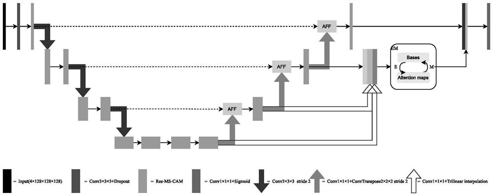
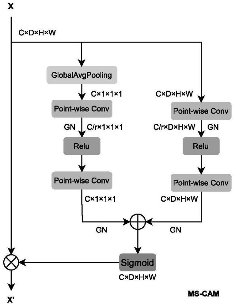
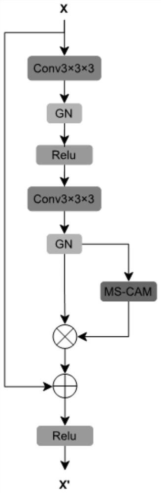
[0062] The following describes in detail the embodiments of the present invention, examples of which are illustrated in the accompanying drawings, wherein the same or similar reference numerals refer to the same or similar elements or elements having the same or similar functions throughout. The embodiments described below with reference to the accompanying drawings are exemplary, only used to explain the present invention, and should not be construed as a limitation of the present invention. The numbers of the steps in the following embodiments are set only for the convenience of description, and the sequence between the steps is not limited in any way, and the execution sequence of each step in the embodiments can be adapted according to the understanding of those skilled in the art Sexual adjustment.

[0063] In the description of the present invention, it should be understood that the orientations related to the description, such as up, down, front, rear, left, right, etc....


PUM


Abstract

The invention discloses a brain tumor image segmentation method, system and device and a storage medium. The method comprises the following steps: carrying out preprocessing and data amplification on a brain tumor image and a label; performing convolution and down-sampling on the brain tumor image, and extracting context semantic information in the brain tumor image to obtain a feature map; performing up-sampling on the feature map, and performing feature fusion on the up-sampled map and the features in the encoder module at the same level; aggregating the feature maps through a feature pyramid fusion module, and inputting the aggregated feature maps into an expectation maximization self-attention module to learn global context information; and aggregating the features and the maximum-level feature map to obtain a final semantic segmentation result. Based on a multi-scale channel attention mechanism, feature extraction and feature fusion, global context information is extracted by adopting a feature pyramid and an expectation maximization attention mechanism, the semantic segmentation precision is improved, and the method can be widely applied to the field of computer vision and image processing.

Description

technical field [0001] The present invention relates to the fields of computer vision and image processing, and in particular, to a method, system, device and storage medium for segmentation of brain tumor images. Background technique [0002] Brain tumor is an abnormal tissue caused by cancerous proliferation of cells caused by uncontrollable factors. According to its origin, it can be divided into primary brain tumor and secondary brain tumor. Primary brain tumor originates from brain cells, and secondary brain tumors Brain tumors spread from tumors that have grown in other organs, distant organs, or adjacent tissues. Gliomas are among the most common primary brain tumors and arise from astrocytes that form the structural backbone of the brain. Based on tumor appearance, gliomas can be divided into four categories I-IV, I and II belong to low-grade gliomas (Low-Grade-Gliomas, LGG), and III and IV belong to high-grade gliomas (High-grade gliomas). Grade-Gliomas, HGG). Ac...

Claims


Application Information

Patent Timeline
IPC IPC(8): G06V10/26G06V10/80G06V10/82G06K9/62G06N3/04G06N3/08
CPCG06N3/08G06N3/048G06N3/045G06F18/253
Inventor 史景伦陈学斌熊静远吕龙飞王鹏
Owner SOUTH CHINA UNIV OF TECH
Why Eureka
Try Eureka