
Angiocardiography device for the department of cardiology

A cardiovascular and cardiology technology, applied in the field of cardiovascular imaging devices in cardiology, can solve the problems of fixed imaging trajectory, low image definition of single-screen imaging device, and difficulty in adapting to disease detection in special populations, so as to improve positioning accuracy, expand The effect of single angiography area and maintaining angiography clarity

Inactive Publication Date: 2022-01-28
HEBI CITY PEOPLES HOSPITAL
View PDF0 Cites 0 Cited by
  • Summary
  • Abstract
  • Description
  • Claims
  • Application Information

AI Technical Summary

Problems solved by technology

[0002] Peritoneal angiography is a method of detection of celiac vascular disease, which injects a contrast agent into the blood vessel, and then uses X-rays to collect blood vessel pictures; based on the fact that X-rays cannot penetrate the contrast agent, the image displayed by the contrast agent under X-ray Therefore, abdominal angiography can accurately reflect the location and degree of celiac vascular disease, thereby obtaining diagnostic information of vascular disease. The image definition of single-screen imaging equipment is low, and its imaging trajectory is relatively fixed, so it is difficult to adapt to the detection of diseases in special populations

Method used


Image

  • Angiocardiography device for the department of cardiology
  • Angiocardiography device for the department of cardiology
  • Angiocardiography device for the department of cardiology

Examples


Embodiment Construction

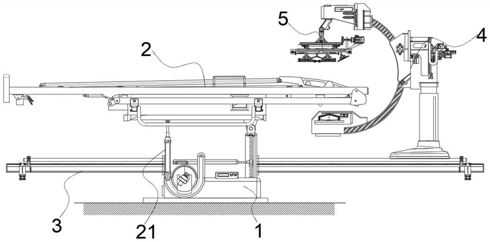
[0045] see figure 1 , in an embodiment of the present invention, a cardiovascular angiography device in the Department of Cardiology, which includes:

[0046] support base 1;

[0047] The receiving frame 2 is horizontally hinged on the support base 1 through the outer strut;

[0048] The hydraulic telescopic rod 21 is hinged on the end of the support base 1 away from the outer pole, and the output end of the hydraulic telescopic rod 21 is connected to the receiving frame 2;

[0049] The outer rail 3 is wound around the support base 1;

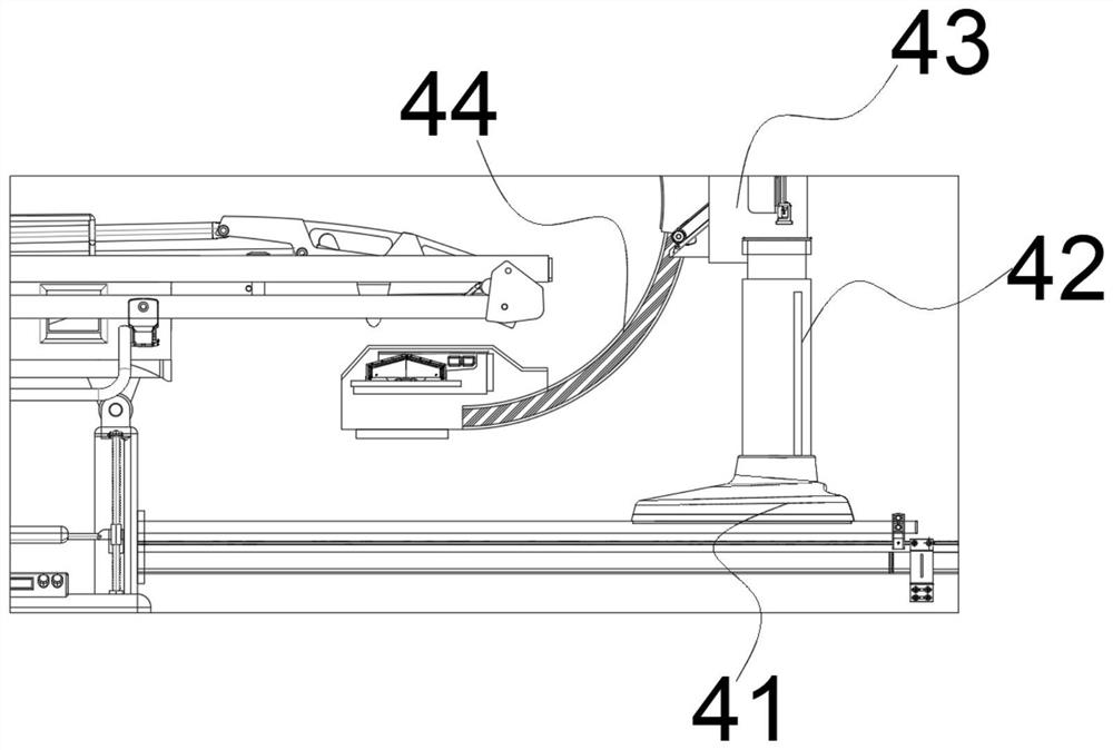
[0050] The sliding bracket 4 is relatively slidable and vertically arranged on the outer rail 3; and

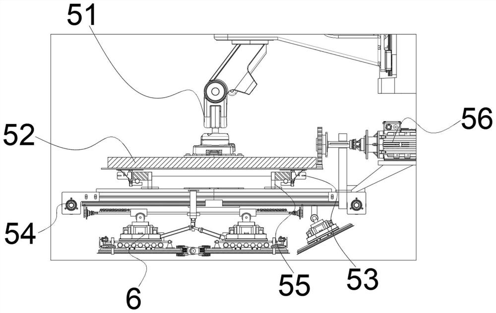
[0051] The radiography adjustment assembly 5 is installed on one end of the sliding bracket 4 and is located above the receiving frame 2, wherein the patient is placed on a flat bed through the receiving frame, and the receiving frame can be driven to deflect by the expansion and contraction of the hydraulic telescopic rod. Thereby changin...


PUM


Abstract

The invention discloses an angiocardiography device for the department of cardiology. The angiocardiography device comprises a supporting base, a bearing frame, a hydraulic telescopic rod, an outer rail part , a sliding support, and aradiography adjusting assembly ; the bearing frame is transversely hinged to the supporting base through an outer support rod; the hydraulic telescopic rod is hinged to the end, away from the outer support rod, of the supporting base, and the output end of the hydraulic telescopic rod is connected with the bearing frame; the outer rail part is wound outside the supporting base; the sliding support is vertically arranged on the outer rail part in a relatively sliding manner; and the radiography adjusting assembly is installed at one end of the sliding support and located above the bearing frame.

Description

technical field [0001] The invention belongs to the technical field of medical equipment, in particular to a cardiovascular angiography device in the department of cardiology. Background technique [0002] Peritoneal angiography is a method of detection of celiac vascular disease, which injects a contrast agent into the blood vessel, and then uses X-rays to collect blood vessel pictures; based on the fact that X-rays cannot penetrate the contrast agent, the image displayed by the contrast agent under X-ray Diagnose celiac vascular lesions, so abdominal angiography can accurately reflect the location and degree of celiac vascular lesions, thereby obtaining diagnostic information for vascular lesions. The image definition of single-screen imaging equipment is low, and its imaging trajectory is relatively fixed, so it is difficult to adapt to the detection of diseases in special populations. Therefore, those skilled in the art provide a cardiovascular angiography device for th...

Claims


Application Information

Patent Timeline
IPC IPC(8): A61B6/04A61B6/00
CPCA61B6/0487A61B6/0407A61B6/481A61B6/504
Inventor 高星王振侯艳艳
Owner HEBI CITY PEOPLES HOSPITAL
Learn More