Clinical test and analysis equipment for endocrine tumor samples and working method of clinical test and analysis equipment

A technology for testing and analyzing endocrine tumors, applied in the field of microscopy, can solve problems such as low efficiency, time-consuming, and inconvenience, and achieve the effect of improving efficiency

Inactive Publication Date: 2021-10-29
QINGDAO MUNICIPAL HOSPITAL
View PDF0 Cites 0 Cited by
  • Summary
  • Abstract
  • Description
  • Claims
  • Application Information

AI Technical Summary

Problems solved by technology

[0003] Clinically, when testing and analyzing endocrine tumor samples, microscopes are used to observe the morphology of endocrine tumor samples. In reality, a patient often has multiple slices, but traditional microscopes can only observe slices at a time. For one slice, it is necessary to remove the previous slice and reinstall the next slice, and when installing the slice, it takes time to calibrate the position of the slice so that the center of the slice is directly facing the center of the light transmission hole. The steps are cumbersome and the efficiency is high. Secondly, when adjusting the mirror to find a bright field of view, the traditional microscope has certain requirements on the external light conditions, and when the microscope is limited by the light conditions and cannot find a bright field of view, the only way to find a bright field of view is to move the microscope , very inconvenient

Method used


Image

  • Clinical test and analysis equipment for endocrine tumor samples and working method of clinical test and analysis equipment
  • Clinical test and analysis equipment for endocrine tumor samples and working method of clinical test and analysis equipment
  • Clinical test and analysis equipment for endocrine tumor samples and working method of clinical test and analysis equipment

Examples


Embodiment 1

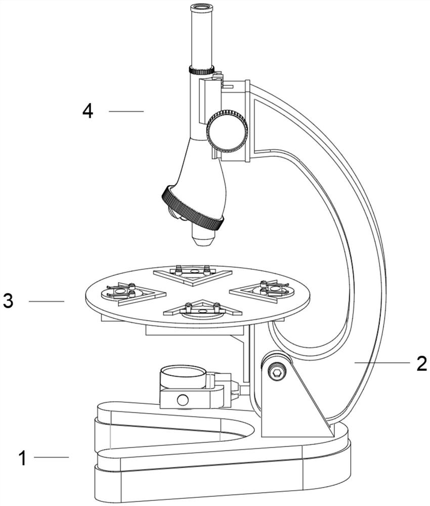
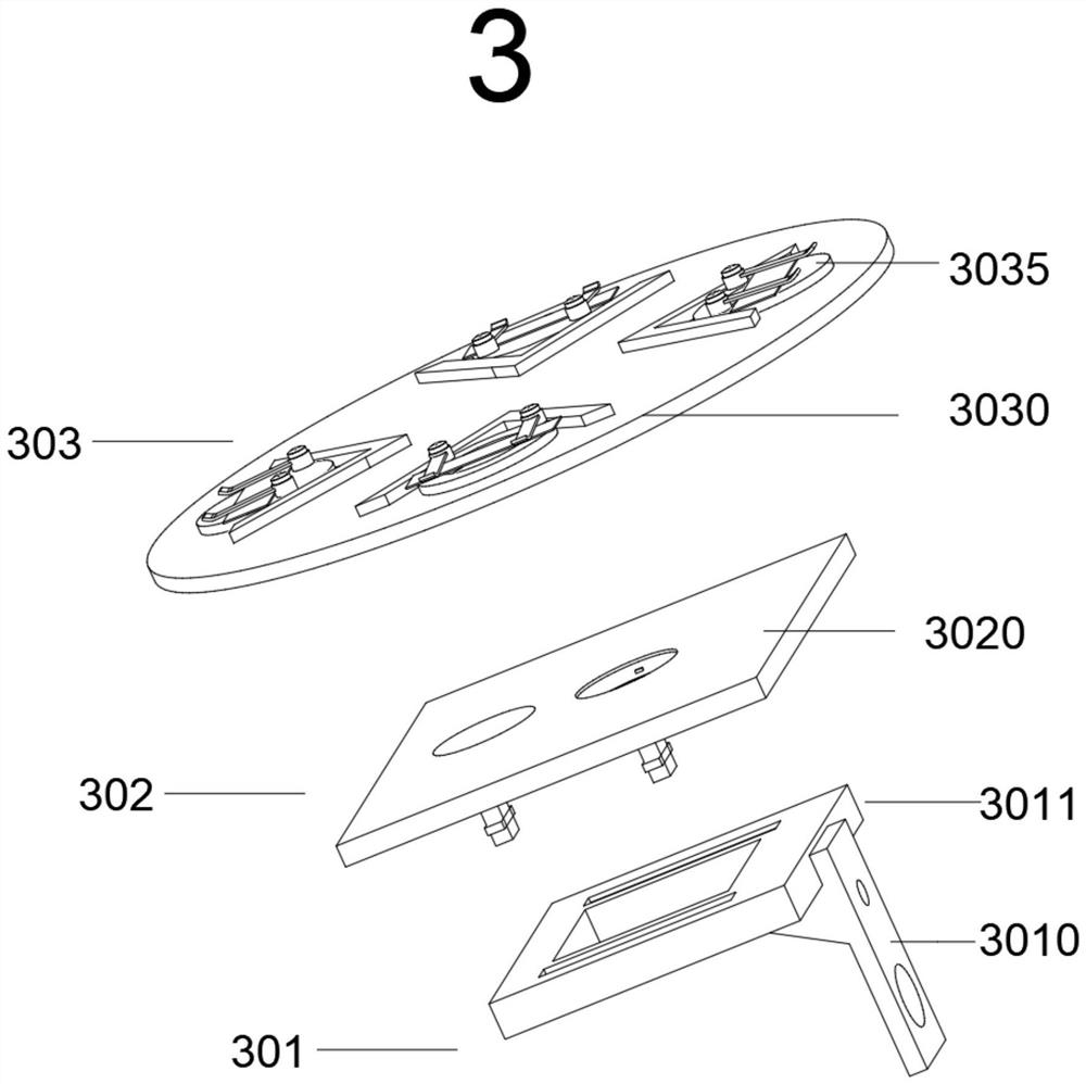
[0040] Such as Figure 1-8 As shown, the embodiment of the present invention provides a clinical test and analysis device for endocrine tumor samples, including a mirror base 1, a mirror arm 2, an object loading device 3 and a lens barrel 4, and the object loading device 3 consists of a first object loading assembly 301, The second loading assembly 302 and the third loading assembly 303 are composed, wherein,

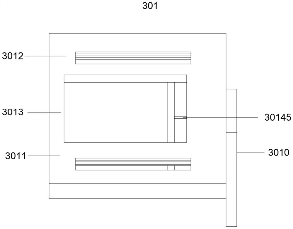
[0041] The first loading assembly 301 includes a support frame 3010, one side of the support frame 3010 is connected to one end of the mirror arm 2, the top of the support frame 3010 is provided with a first object stage 3011, and one side of the surface of the first object stage 3011 is opened There is a through groove 3013, and both sides of the through groove 3013 are provided with chute 3012, and the bottom of the first stage 3011 is provided with a driving device 3014 near the side of the support frame 3010, and the driving device 3014 is used to push the slide bar...

Embodiment 2

[0061] A working method of an endocrine tumor sample clinical test analysis device, comprising the following steps:

[0062] S1, adjust the loading device 3 to find a bright field of view: install the eyepiece and the objective lens on both ends of the lens barrel 4 in sequence, turn the knob 30147, and the driving device 3014 pushes the slider 3025 to slide inside the chute 3012 through the slider 3015, so that The through slot 3013, the first light transmission hole 3024, the second light transmission hole 3034 and the third light transmission hole 30352 are connected to each other. Adjust the reflector to find a bright field of view. When the light conditions are limited, the adjustment of the reflector cannot find a bright field of view. , continue to rotate the knob 30147, so that the first light transmission hole 3024, the second light transmission hole 3034 and the third light transmission hole 30352 continue to move within the range of the through groove 3013, then adju...


PUM


Abstract

The invention relates to clinical test and analysis equipment for endocrine tumor samples and a working method thereof. The equipment comprises a lens base, a lens arm, a loading device and a lens cone, wherein the loading device is composed of a first loading assembly, a second loading assembly and a third loading assembly, the third loading assembly is rotatably connected with the second loading assembly, and a plurality of limiting assemblies are arranged on the surface of a third loading table. The limiting assembly is used for installing the slices, so that the device can install a plurality of slices at the same time, the positions of the slices can be fixed by placing the patient slices on the surface of the limiting assembly, after one slice is observed and recorded, the third objective table is rotated until the third objective table is fixed again, and at the moment, the other limiting assembly just arrives at the position where the previous limiting assembly is located, a slice fixed to the surface of the limiting assembly can be observed only by adjusting the fine focusing screw, repeated slice installation and repeated focusing are not needed, the operation steps are simplified, and the slice observation efficiency is improved.

Description

technical field [0001] The invention relates to the technical field of microscopes, in particular to a clinical testing and analysis device for endocrine tumor samples and a working method thereof. Background technique [0002] Neuroendocrine tumors are tumors that arise from neuroendocrine cells. Neuroendocrine cells are a large class of cells that have a neuroendocrine phenotype in the body and can produce a variety of hormones. Neuroendocrine cells are distributed throughout the body, so neuroendocrine tumors can occur anywhere in the body, but the most common are stomach, intestinal Neuroendocrine tumors of the digestive system, such as pancreas and pancreas, account for about 2 / 3 of all neuroendocrine tumors. The incidence of neuroendocrine tumors in European and American populations is 2.5-5 per 100,000 people, and the incidence has increased by 5 times in the past 30 years. Compared with other tumors, the incidence of neuroendocrine tumors has increased more rapidly....

Claims


Application Information

Patent Timeline
Patent Type & Authority Applications(China)
IPC IPC(8): G01N21/84G01N21/01G01N21/13
CPCG01N21/84G01N21/01G01N21/13
Inventor 贾少丹李靖肖芝秀
Owner QINGDAO MUNICIPAL HOSPITAL
Why Eureka
Try Eureka