
Medical image processing method and device, medium and electronic equipment

A medical image and processing method technology, applied in the field of image processing, can solve the problem of inability to distinguish between arteries and veins

Pending Publication Date: 2021-08-17
SHANGHAI CHANGZHENG HOSPITAL +1
View PDF0 Cites 1 Cited by
  • Summary
  • Abstract
  • Description
  • Claims
  • Application Information

AI Technical Summary

Problems solved by technology

[0003] In view of the above-mentioned shortcomings of the prior art, the purpose of the present invention is to provide a medical image processing method, device, medium and electronic equipment, which are used to solve the problem that arteries and veins in lung medical images cannot be accurately processed in the prior art. the problem of distinguishing

Method used


Image

  • Medical image processing method and device, medium and electronic equipment
  • Medical image processing method and device, medium and electronic equipment
  • Medical image processing method and device, medium and electronic equipment

Examples


Embodiment Construction

[0058] Embodiments of the present invention are described below through specific examples, and those skilled in the art can easily understand other advantages and effects of the present invention from the content disclosed in this specification. The present invention can also be implemented or applied through other different specific implementation modes, and various modifications or changes can be made to the details in this specification based on different viewpoints and applications without departing from the spirit of the present invention. It should be noted that, in the case of no conflict, the following embodiments and features in the embodiments can be combined with each other.

[0059] It should be noted that the diagrams provided in the following embodiments are only schematically illustrating the basic ideas of the present invention, and only the components related to the present invention are shown in the diagrams rather than the number, shape and size of the compon...


PUM


Abstract

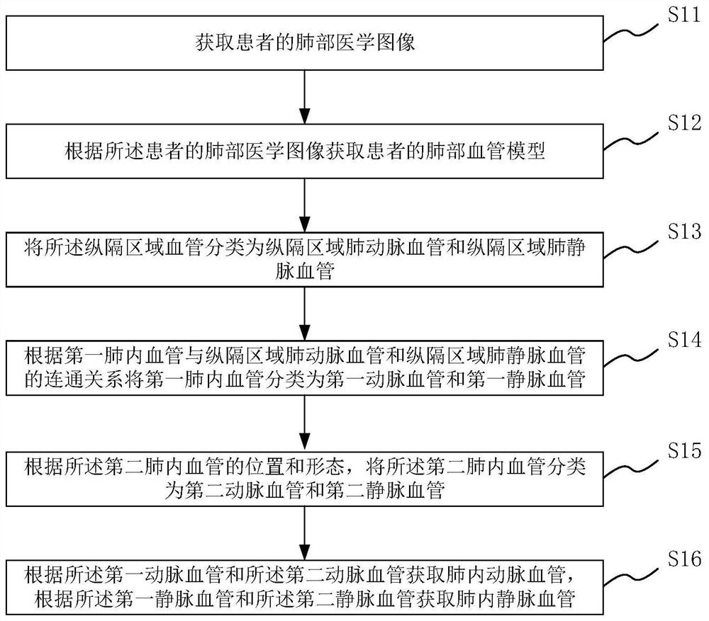
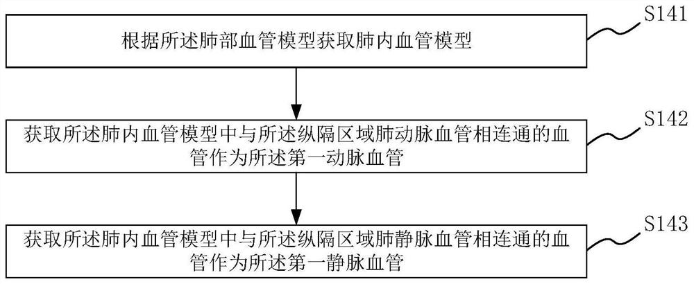
The invention provides a medical image processing method and device, a medium and electronic equipment. The medical image processing method comprises the following steps: acquiring a lung medical image of a patient; acquiring a lung vessel model of the patient according to the lung medical image of the patient; classifying the mediastinal region blood vessels into mediastinal region pulmonary artery blood vessels and mediastinal region pulmonary vein blood vessels; classifying the first intra-pulmonary blood vessels into first artery blood vessels and first vein blood vessels according to the communication relationship between the first intra-pulmonary blood vessels and the mediastinal region pulmonary artery blood vessels and the mediastinal region pulmonary vein blood vessels; classifying the second intra-pulmonary blood vessels into second artery blood vessels and second vein blood vessels according to the positions and shapes of the second intra-pulmonary blood vessels; according to the first artery blood vessel and the second artery blood vessel, obtaining an intra-pulmonary artery blood vessel, and according to the first vein blood vessel and the second vein blood vessel, obtaining an intra-pulmonary vein blood vessel. The medical image processing method can accurately distinguish the vein and the artery in the lung medical image.

Description

technical field [0001] The invention relates to an image processing method, in particular to a medical image processing method, device, medium and electronic equipment. Background technique [0002] With the development of medical imaging technology and the application of computer image processing technology in the field of medical image processing, medical personnel can use computer image processing technology to analyze and process medical images to obtain auxiliary diagnosis conclusions for related diseases. For some lung diseases, it is necessary to distinguish arteries and veins in lung medical images before obtaining auxiliary diagnosis conclusions. However, the inventors have found in practical applications that the existing image segmentation technology can only segment medical images of the lungs to obtain pulmonary blood vessels, but cannot accurately distinguish arteries and veins in the medical images of the lungs. Contents of the invention [0003] In view of...

Claims


Application Information

Patent Timeline
IPC IPC(8): A61B6/03G06N3/08G06T7/11
CPCA61B6/032A61B6/50A61B6/5211G06T7/11G06N3/08G06T2207/30061G06T2207/20221G06T2207/30101
Inventor 刘士远范丽蒲瑜周秀秀夏艺管宇张迪涂文婷张凡徐攀龙杨华傅琪钲路洋房劬
Owner SHANGHAI CHANGZHENG HOSPITAL
Learn More