
Angiography device and method

A technology for angiography and mounting chambers, which is applied in the field of medical appliances, can solve the problems of medical staff inconvenience, calculation errors, prolonging the inspection time, etc., and achieve the effects of convenient operation, convenient fixation, and flexible use

Active Publication Date: 2021-07-13
XIEHE HOSPITAL ATTACHED TO TONGJI MEDICAL COLLEGE HUAZHONG SCI & TECH UNIV
View PDF17 Cites 0 Cited by
  • Summary
  • Abstract
  • Description
  • Claims
  • Application Information

AI Technical Summary

Problems solved by technology

[0003] When performing cerebral angiography on a patient, the patient needs to lie flat on the angiography table with the head fixed. For patients who are nervous or the angiography time is too long, it is easy to relax their vigilance and inadvertently change the head Therefore, it affects the effect of cerebral angiography, causes calculation errors, prolongs the inspection time, and brings inconvenience to medical staff. Therefore, it is necessary to fix the patient's head with a specific device
[0004] However, the fixing devices in the prior art are generally installed on the radiography bed, which is not very convenient to fix the patient, and the fixing method is cumbersome, and the fixing of the patient cannot be quickly released when an accident occurs.

Method used


Image

  • Angiography device and method
  • Angiography device and method
  • Angiography device and method

Examples


Embodiment Construction

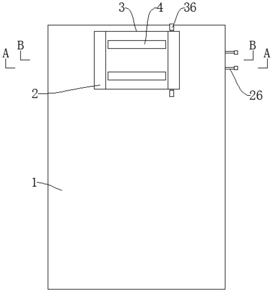
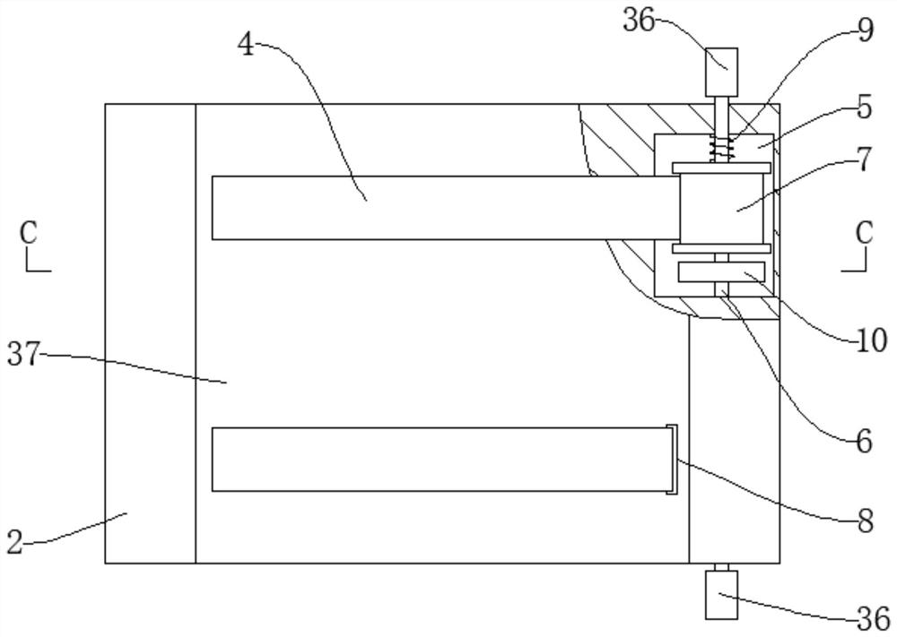
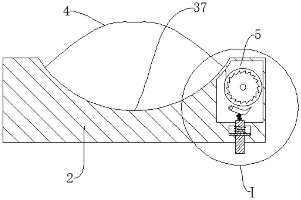
[0032] Such as Figure 1 to Figure 8 As shown, an angiography apparatus includes a bed body 1 on which a headrest 2 is detachably mounted, and a positioning groove 3 matching the headrest 2 is provided on the bed body 1, so that A locking mechanism is provided between the bed body 1 and the headrest 2; two sets of fixing mechanisms are arranged up and down on the headrest 2, and the fixing mechanism includes a fixing belt 4, the left end of which is fixed Connected to the left end of the headrest 2, the right end of the headrest 2 is provided with an installation cavity 5, and the installation cavity 5 is rotatably installed with an installation shaft 6 extending forward and backward, and the installation shaft 6 is coaxially fixed with a A winding wheel 7, the right end of the fixing belt 4 is wound on the winding wheel 7, and the headrest 2 is provided with a port 8 for the fixing belt 4 to enter the installation cavity 5, so Between the installation shaft 6 and the side wa...


PUM


Abstract

The invention relates to an angiography device and method. A headrest is detachably installed on a bed body, a locking mechanism is arranged between the bed body and the headrest, the right end of a fixing belt is wound on a winding wheel, a torsional spring used for enabling the winding wheel to wind the fixing belt is connected between an installation shaft and the side wall of an installation cavity, a ratchet wheel is further coaxially and fixedly installed on the installation shaft, a first spring is connected between the upper end of the movable column and the pawl, a second spring is connected between the baffle and the top wall of the auxiliary cavity, and a linkage mechanism used for driving the pressing column to move up and down is arranged in the positioning groove. The headrest and the bed body are detachable, the headrest is fixed to the head of a patient, after the position of the headrest is adjusted till the patient feels comfortable, the headrest and the bed body are fixed, fixing is convenient, and the comfort degree is high; the shifting handle is shifted to drive the bolt to be separated from the insertion hole, so that the limitation on the insertion block can be relieved, then the headrest can be separated from the bed body immediately, and the fixation on the head of the patient is relieved.

Description

technical field [0001] The invention relates to the technical field of medical appliances, in particular to an angiography device and method. Background technique [0002] Cardiovascular and cerebrovascular diseases mostly occur in middle-aged and elderly people, with high morbidity, mortality and disability rates. With the acceleration of the aging society, cardiovascular and cerebrovascular diseases have become the number one killer threatening human health. Cardiovascular and cerebrovascular diseases Contrast examination is widely used in clinical practice due to its technical characteristics of less trauma, quick detection and relatively obvious results. [0003] When performing cerebral angiography on a patient, the patient needs to lie flat on the angiography table with the head fixed. For patients who are nervous or the angiography time is too long, it is easy to relax their vigilance and inadvertently change the head Therefore, it affects the effect of cerebral angi...

Claims


Application Information

Patent Timeline
Patent Type & Authority Applications(China)
IPC IPC(8): A61B6/04
CPCA61B6/0421A61B6/504
Inventor 任衍乔郑传胜陈磊阚雪锋
Owner XIEHE HOSPITAL ATTACHED TO TONGJI MEDICAL COLLEGE HUAZHONG SCI & TECH UNIV
Learn More