
Tumor sampling device facilitating tumor separation

A sampling device and tumor technology, applied in the field of medical devices, can solve the problems of poor tool stability, hand scratches, low safety, etc., and achieve the effects of reducing hand scratches, low manufacturing cost, and convenient operation.

Active Publication Date: 2021-07-02
陕西省肿瘤医院
View PDF5 Cites 3 Cited by
  • Summary
  • Abstract
  • Description
  • Claims
  • Application Information

AI Technical Summary

Problems solved by technology

[0002] Tumor is a new organism formed by the abnormal proliferation of local tissue cells under the action of various tumorigenic factors, because this new organism is mostly in the form of space-occupying blocky protrusions, also known as neoplasms, tumors are used in the current medical treatment There are still great difficulties in the field, and there is still a long way to go. Surgical resection is the best treatment for early tumors. After tumor resection, in order to classify tumors by pathological subtypes, it is often necessary Due to the different shapes of tumors, some tumor masses have a smooth surface, or there are calcified tissues inside, the suction by needle tube has great limitations in the process of sampling, which often requires a scalpel to treat the tumor. Then, a certain volume of internal tissue is taken out for further analysis and confirmation. During this process, due to the different degrees of softness and hardness inside or on the surface of the tumor block, sliding often occurs, resulting in poor stability of the knife during the sampling process and easy cutting. There is a certain risk in the hands of doctors, so it is necessary to install a tumor sampling device that is convenient for separating tumors to solve these problems, such as: when sampling with a knife cutting method, it is easy to slip, the stability is not good, and there are hand scratches risk, low security

Method used


Image

  • Tumor sampling device facilitating tumor separation
  • Tumor sampling device facilitating tumor separation
  • Tumor sampling device facilitating tumor separation

Examples


Embodiment 1

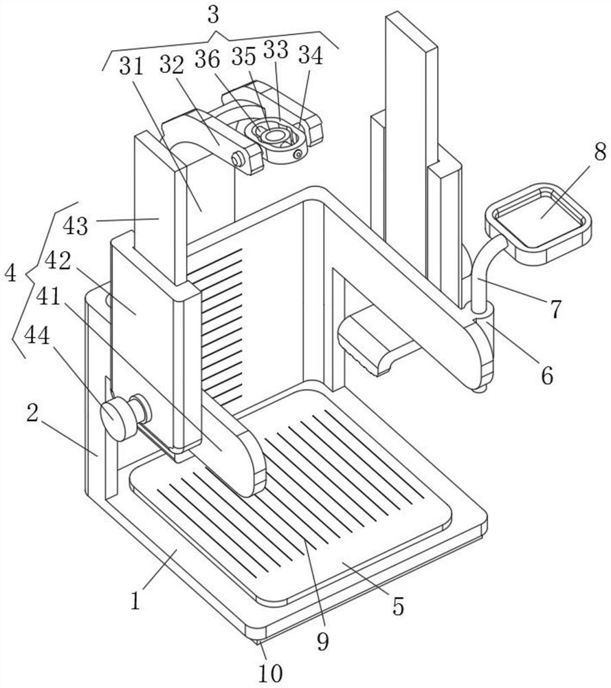
[0014] Such as Figure 1 to Figure 3 As shown, the present invention discloses a tumor sampling device for conveniently separating tumors. The adopted technical solution includes a mounting plate 1, a vertical plate 2, a limit mechanism 3 and a positioning mechanism 4, and the position of the rear edge of the mounting plate 1 is The vertical plate 2 is fixedly connected on the surface, and the front side of the vertical plate 2 is connected with a positioning mechanism 4. The limit mechanism 3 includes a support plate 31, a connecting head 32, a first connecting ring 33, a first connecting shaft 34, a second The connecting ring 35 and the second connecting shaft 36, the supporting plate 31 are fixedly connected on the surface of the middle position of the rear side of the vertical plate 2, and the upper end of the vertical plate 2 is fixedly connected with a forwardly curved connecting head 32, the front end of the connecting head 32 It is U-shaped, the first connecting ring 3...


PUM


Abstract

A tumor sampling device facilitating tumor separation, disclosed in the invention, comprises a mounting plate, a vertical plate, a limiting mechanism and a positioning mechanism, the vertical plate is fixedly connected to the surface of the edge of the rear side of the mounting plate, and the positioning mechanism is connected to the front side of the vertical plate; the limiting mechanism comprises a supporting plate, a connector, a first connecting ring, a first connecting shaft, a second connecting ring and a second connecting shaft, the supporting plate is fixedly connected to the surface of the middle position of the rear side of the vertical plate, the connector bent forwards is fixedly connected to the upper end of the vertical plate, and the front end of the connector is in a U shape; and the first connecting ring is oval, first connecting shafts are fixedly connected to the surfaces of the two sides of the first connecting ring, and the first connecting shafts are rotationally connected with the two sides of the U-shaped end of the connector. The tumor sampling device facilitating tumor separation can support a surgical knife, is good in stability, reduces the cutting risk and is good in safety.

Description

technical field [0001] The invention relates to the technical field of medical devices, in particular to a tumor sampling device for conveniently separating tumors. Background technique [0002] Tumor is a new organism formed by the abnormal proliferation of local tissue cells under the action of various tumorigenic factors, because this new organism is mostly in the form of space-occupying blocky protrusions, also known as neoplasms, tumors are used in the current medical treatment There are still great difficulties in the field, and there is still a long way to go. Surgical resection is the best treatment for early tumors. After tumor resection, in order to classify tumors by pathological subtypes, it is often necessary Due to the different shapes of tumors, some tumor masses have a smooth surface, or there are calcified tissues inside, the suction by needle tube has great limitations in the process of sampling, which often requires a scalpel to treat the tumor. Then, a c...

Claims


Application Information

Patent Timeline
Patent Type & Authority Applications(China)
IPC IPC(8): G01N1/04G01B5/02
CPCG01N1/04G01B5/02
Inventor 孙海凤杨建军郭亚焕
Owner 陕西省肿瘤医院
Learn More