Cell image segmentation method

An image and cell technology, applied in the field of cell analysis, can solve the problems of difficult cell observation and labeling, difficult for researchers to observe for a long time, and inability to accurately segment real cell edges, etc., to improve cell adhesion and eliminate cells. Boundary aperture, the effect of enriching target detail information

Active Publication Date: 2020-12-08
KUNMING UNIV OF SCI & TECH +1
View PDF6 Cites 1 Cited by
  • Summary
  • Abstract
  • Description
  • Claims
  • Application Information

AI Technical Summary

Problems solved by technology

[0004]1. The number of cells is huge and densely distributed, requiring huge human resources;
[0005]2. Due to the microscopic characteristics of cells, it is difficult for researchers to observe for a long time, and the observation process is very subjective, so it is difficult to ensure the correctness of the observation results;
[0006]3. The growth environment of stem cells is an important experimental parameter. The dye labeling method used in traditional cell observation methods will affect the activity of cells, which makes the cells It is more difficult to observe and mark the
At the same time, for the segmentation of adhesive stem cell images with apertures, the traditional threshold method cannot accurately obtain the pixel distribution information of the cells and the background in the image, resulting in the inability to accurately segment the edges of the real cells, and the segmentation effect is not good; and the principle of phase contrast microscopy is used to eliminate the influence of the aperture , requiring a complex modeling process

Method used


Image

  • Cell image segmentation method
  • Cell image segmentation method
  • Cell image segmentation method

Examples


Embodiment 1

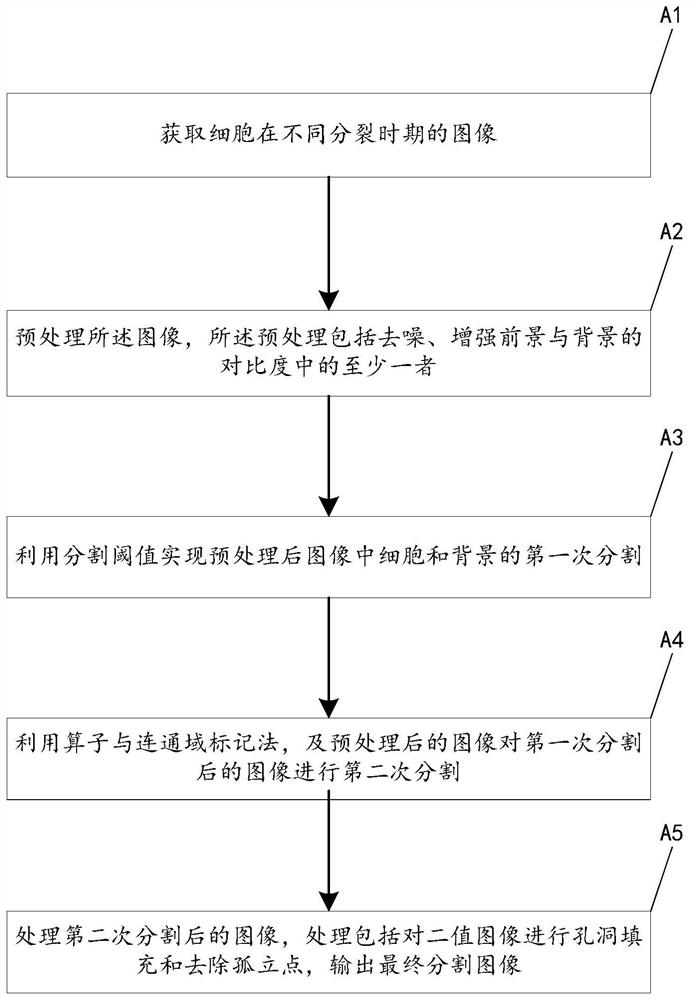
[0038] figure 1 The flow chart of the cell image segmentation method of the embodiment of the present invention is given, such as figure 1 As shown, the segmentation method of the cell image comprises the following steps:

[0039] (A1) Using an aberration microscope to obtain images of cells at different stages of division;

[0040] (A2) preprocessing the image, the preprocessing includes at least one of denoising and enhancing the contrast between the foreground and the background; image denoising and contrast enhancement are prior art in this field;

[0041] (A3) utilizing the segmentation threshold to realize the first segmentation of cells and background in the preprocessed image; the segmentation threshold is;

[0042] Obtain the pixel intensity peak m and pixel growth rate s of the preprocessed image;

[0043] get initial threshold R represents the dynamic deviation value, k is a positive constant;

[0044] If the threshold Then return to continue to calculate th...

Embodiment 2

[0063] An application example of the cell image segmentation method according to Embodiment 1 of the present invention in bone marrow stem cells.

[0064] The segmentation method of the bone marrow stem cell image in this application example, such as figure 1 As shown, the segmentation method of the bone marrow stem cell image comprises the following steps:

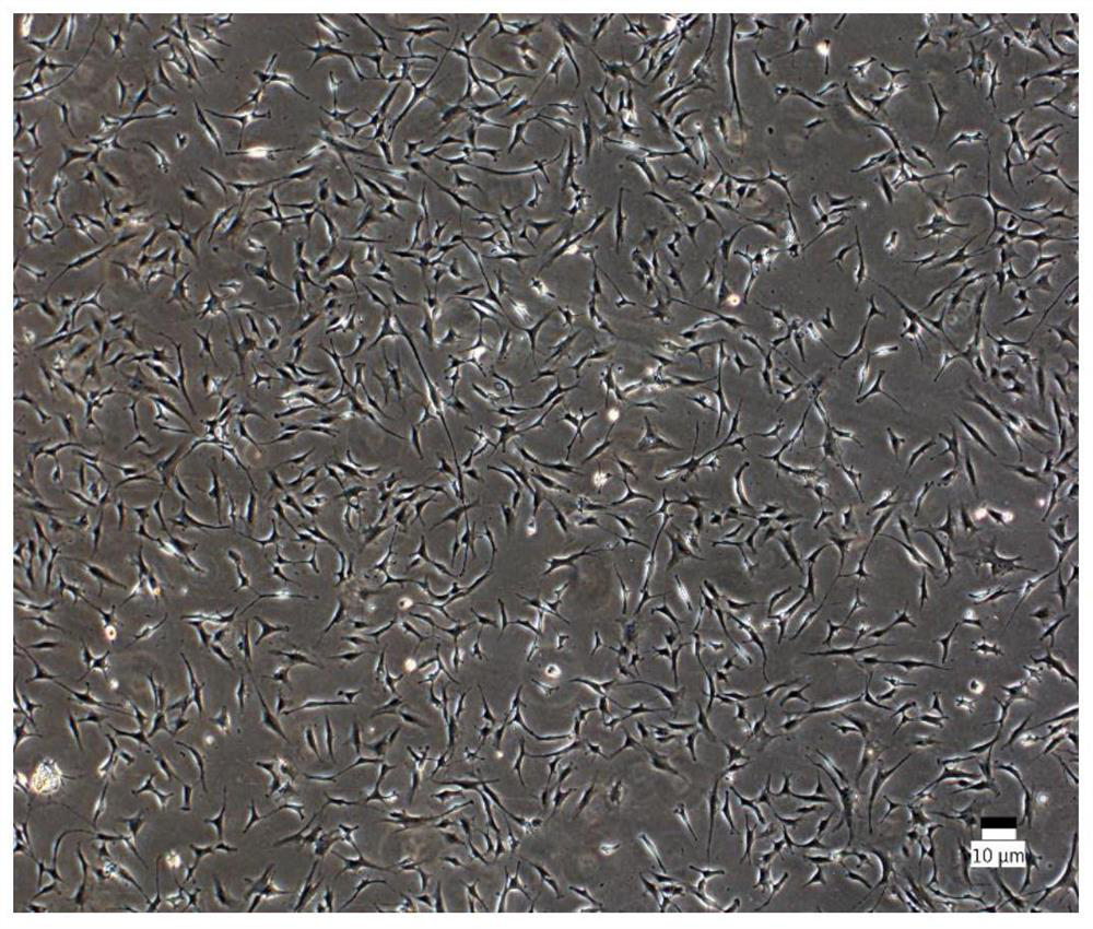
[0065] (A1) Using aberration microscopy to obtain images of cells at different stages of division, such as figure 2 shown;

[0066] (A2) preprocessing the image, the preprocessing includes denoising and enhancing the contrast between the foreground and the background, specifically;

[0067] Using a Gaussian filter to denoise is very effective for suppressing noise that obeys a normal distribution;

[0068] Perform morphological top-hat transformation and bottom-hat transformation on the image, and calculate the top-hat transformation and bottom-hat transformation,

[0069] T bat (I)=(I·b)-I; Indicates the openin...


PUM


Abstract

The invention provides a cell image segmentation method. The cell image segmentation method comprises the following steps: (A1) acquiring an image of a cell in a division period; (A2) preprocessing the image, wherein the preprocessing comprises at least one of denoising and enhancing the contrast ratio of a foreground and a background; (A3) utilizing a segmentation threshold to realize first segmentation of cells and a background in the preprocessed image; (A4) carrying out second segmentation on the image after the first segmentation by utilizing an operator, a connected domain marking methodand the preprocessed image; and (A5) processing the image after the second segmentation, wherein the processing includes the operation of performing hole filling on the binary image and removing isolated points, and outputting a final segmented image. The method has the advantages of high segmentation precision and the like.

Description

technical field [0001] The invention relates to cell analysis, in particular to a cell image segmentation method. Background technique [0002] Stem cells are cells with the potential of self-replication and multi-directional differentiation. They have the ability to form other cells or tissues in the human body. They are called "universal cells" in the medical field. The large medical application value is of great significance to the maintenance of human health and the continuation of life. [0003] The growth of stem cells is a process of continuous division and differentiation, and random movement. Traditional research methods mainly rely on researchers to understand and analyze the growth status of cells through naked eye observation, which has the following disadvantages: [0004] 1. The number of cells is huge and densely distributed, which requires huge human resources; [0005] 2. Due to the microscopic characteristics of cells, it is difficult for researchers to o...

Claims


Application Information

Patent Timeline
Patent Type & Authority Applications(China)
IPC IPC(8): G06T7/00G06T7/13G06T7/136G06T7/187G06T7/194G06T5/00
CPCG06T7/0012G06T7/13G06T7/136G06T7/187G06T7/194G06T5/002G06T2207/10061G06T2207/30024
Inventor 伏金浩洪欢欢闻路红史振志王家杰张果刘楠楠
Owner KUNMING UNIV OF SCI & TECH
Why Eureka
Try Eureka