
Method for promoting proliferation of right ventricular myocardial cells in vivo

A cardiomyocyte proliferation and cardiomyocyte technology, applied in the field of promoting right ventricular cardiomyocyte proliferation in vivo, can solve problems such as research without right ventricle

Inactive Publication Date: 2020-07-31
SHANGHAI CHILDRENS MEDICAL CENT AFFILIATED TO SHANGHAI JIAOTONG UNIV SCHOOL OF MEDICINE
View PDF0 Cites 0 Cited by
  • Summary
  • Abstract
  • Description
  • Claims
  • Application Information

AI Technical Summary

Problems solved by technology

[0007] 4. The current technology is all aimed at the study of the left ventricle, and there is no relevant report on the study of the right ventricle
[0009] There is no report about a method of the present invention for promoting the proliferation of right ventricular cardiomyocytes in vivo

Method used


Image

  • Method for promoting proliferation of right ventricular myocardial cells in vivo
  • Method for promoting proliferation of right ventricular myocardial cells in vivo
  • Method for promoting proliferation of right ventricular myocardial cells in vivo

Examples


Embodiment 1

[0043] 1 Experimental materials

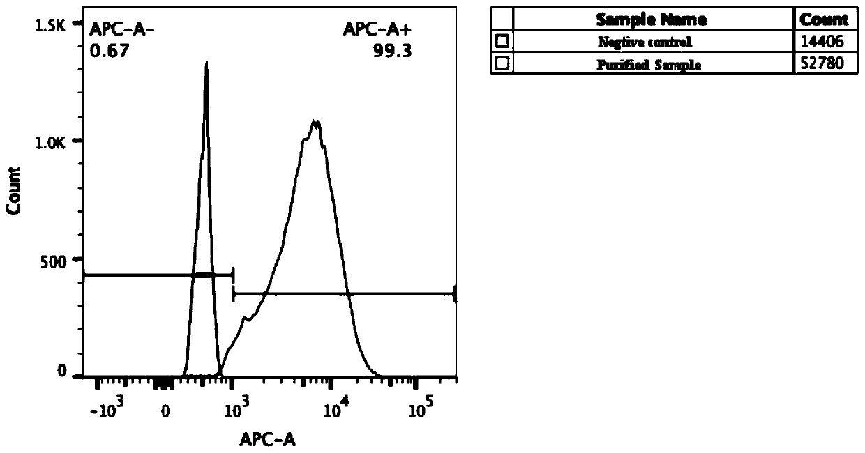
[0044] Monocrotaline (purchased from Sig-ma Company, USA) was prepared into a 1% solution with anhydrous ethanol and normal saline at a ratio of 2:8, mixed evenly, and set aside.

[0045] Thirty male SD neonatal rats (1-3 days after birth) were purchased from Shanghai Second Military Medical University and randomly divided into two groups, namely the experimental group and the control group, with 15 rats in each group.

[0046] Table 1. Reagents

[0047]

[0048] Table 2 Antibodies

[0049]

[0050]

[0051] 2 Experimental methods

[0052] Rats in the experimental group were intraperitoneally injected with a solution of ethanol saline 2:8, and rats in the control group were subcutaneously injected with 50 mg / kg of monocrotaline once at the back of the neck, and were fed for 14 days under the same environment.

[0053] 2.1 After feeding for 14 days, the general condition of the rats was observed, and the pulmonary artery pressure, c...

Embodiment 2

[0068] 1 Experimental method

[0069] Myocardial tissue from the right ventricular outflow tract of tetralogy of Fallot (TOF) younger than 6 months old (source: children from Shanghai Children's Medical Center affiliated to Shanghai Jiaotong University School of Medicine) was selected for detection.

[0070] 2 Experimental results

[0071] It was found that the expression of myocardial tissue proliferation markers was significantly increased in children with elevated pressure load. ( Figure 5 G-K)

[0072] 3 Conclusion

[0073] The above results indicate that elevated pressure load can promote the proliferation of right ventricular cardiomyocytes in human infants.

[0074] The present invention studies a method for proliferation of right ventricular cardiomyocytes in vivo, and experiments prove that promoting the proliferation and regeneration of endogenous cardiomyocytes to replace cardiomyocytes with impaired systolic function can effectively alleviate or reverse the sy...


PUM


Abstract

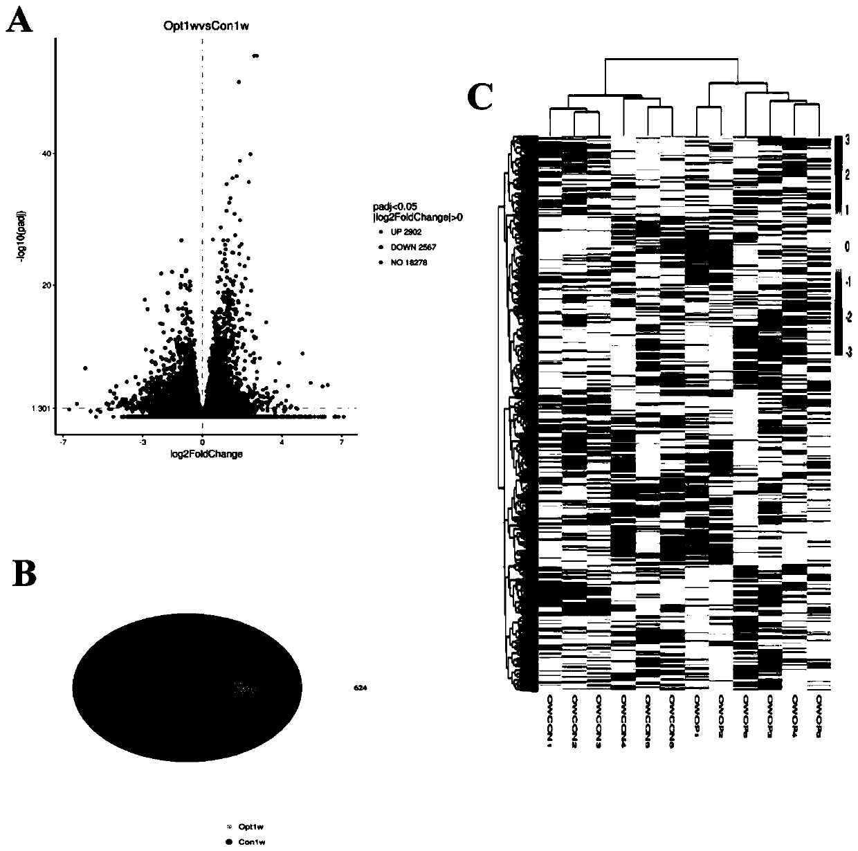
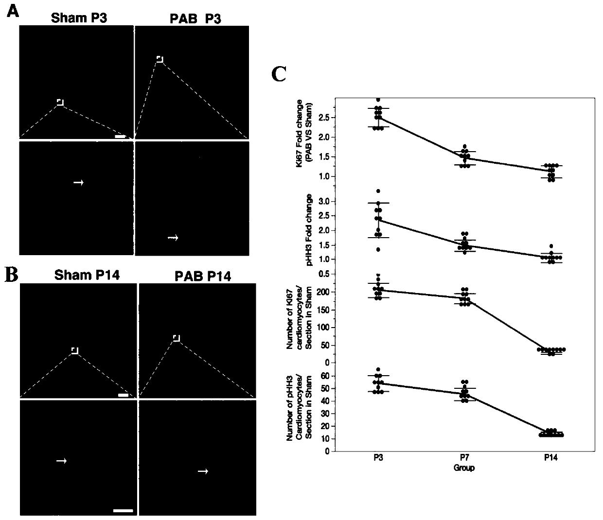
The invention relates to the technical field of biology, in particular to a method for promoting proliferation of right ventricular myocardial cells in vivo. The method comprises the following steps:constructing a pulmonary arterial hypertension animal model by injecting wild lily alkali, and further causing right ventricular pressure rise and right ventricular wall hypertrophy of the model to promote proliferation of the animal right ventricular myocardial cells. The result of the invention also finds that the pressure overload greatly changes the gene expression of myocardial cells, 5469 differential expression genes exist, 2902 genes are up-regulated, 2567 genes are down-regulated, and the smaller the animal age is, the more significant the proliferation marker is. According to the invention, proliferation and regeneration of endogenous myocardial cells are promoted to replace myocardial cells with damaged contractile functions, so that the symptom of heart failure can be effectively relieved or reversed. The method has the advantages that a possible method or means is provided for treatment of heart failure, especially right heart failure, and a mammal model more reliable thanexisting models is provided for heart regeneration research.

Description

technical field [0001] The invention relates to the field of biotechnology, in particular to a method for promoting proliferation of right ventricle cardiomyocytes in vivo. Background technique [0002] At present, the mammalian models used in the study of cardiac regeneration in vivo mainly include neonatal rat myocardial infarction model and apexectomy model. However, these two models are currently controversial - some scholars believe that there is no complete heart regeneration (references. 1.J Mol Cell Cardiol.2016Jan; 90:47-52; 2.Cell Rep.2019Jul 2; 28( 1):190-201.e3; 3.Circulation.2017Sep 19;136(12):1123-1139; 4.J Mol Cell Cardiol.2015Feb;79:315-8.). And RNA sequencing technology proves that in the mouse myocardial infarction model, compared with the sham operation group and the operation group, there are only a very small number of differential genes (294) (Aging (AlbanyNY). 2012Dec; 4(12): 966-77). Furthermore, multi-cell transcriptome analysis technology showed t...

Claims


Application Information

Patent Timeline
Patent Type & Authority Applications(China)
IPC IPC(8): A61K45/00A61K31/407A61P9/00A61P9/04A01K67/02
CPCA01K67/02A01K2227/105A01K2267/0375A61K31/407A61K45/00A61P9/00A61P9/04
Inventor 叶霖财洪海筏刘锦纷仇黎生姜川张海波张浩肖颖颖
Owner SHANGHAI CHILDRENS MEDICAL CENT AFFILIATED TO SHANGHAI JIAOTONG UNIV SCHOOL OF MEDICINE
Learn More