Fluorescent carbon dots for self-targeting cell nucleus and preparation method and application thereof
A technology of fluorescent carbon dots and cell nuclei, applied in fluorescence/phosphorescence, chemical instruments and methods, nano-carbon, etc., to achieve the effects of energy saving, time saving, good solubility and dispersibility, and stable optical properties in the preparation process
- Summary
- Abstract
- Description
- Claims
- Application Information
AI Technical Summary
Problems solved by technology
Method used
Image


Examples
Embodiment 1
[0030] Preparation method of carbon dots:
[0031] 1) Weigh 0.17g of copper chloride and 0.1g of tryptophan into a glass beaker, add 20mL of deionized water, stir thoroughly and ultrasonically dissolve for 15 minutes;
[0032] 2) Transfer the reaction mixture to a hydrothermal reaction kettle, place it in an oven, and react at 180°C for 3 hours;
[0033] 3), after the reaction stops, let it stand and cool to room temperature, filter, remove insoluble matter to obtain a dark yellow solution, and filter with a 0.22 μm filter membrane to obtain a pure aqueous solution of carbon dots;
[0034] 4), freeze-drying the carbon dot aqueous solution to obtain a carbon dot solid.
Embodiment 2
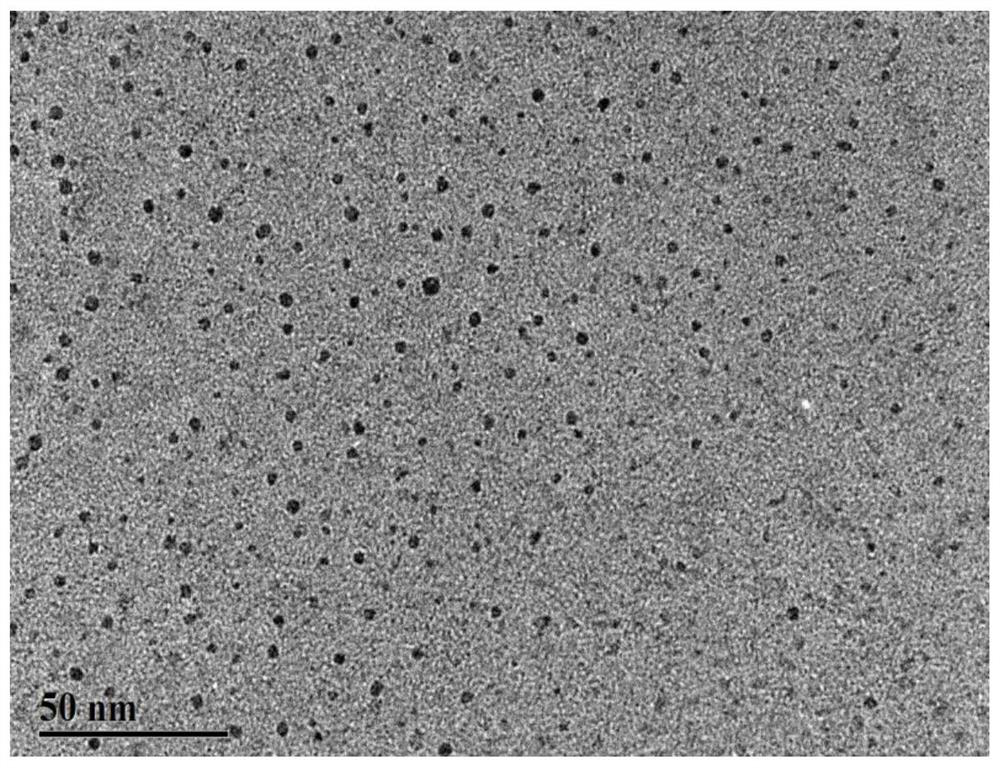
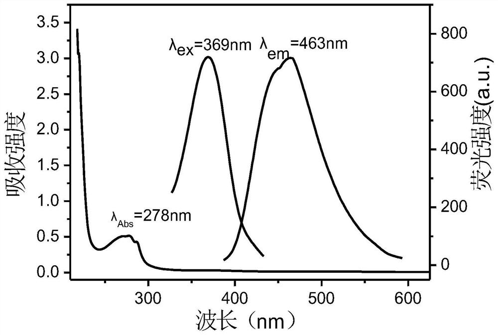
[0036] The fluorescent carbon dot prepared in embodiment 1 is characterized, and its transmission electron microscope picture is as follows figure 1 , the carbon dots present monodisperse spherical particles in morphology, and the particle size is about 4.84nm; the ultraviolet absorption spectrum and fluorescence excitation-emission spectrum of the above-mentioned synthesized carbon dots are as follows: figure 2 , showing that the absorption peak of carbon dots is 278nm, and the optimal excitation wavelength and emission wavelength are 369 and 464nm; the XPS spectrum of the synthesized carbon dots is as follows image 3 , which proves that the main elements contained in carbon dots are carbon, hydrogen, oxygen, nitrogen, copper, and chlorine; the infrared spectrum of the synthesized carbon dots is as follows Figure 4 , indicating that the carbon dot has a benzene ring structure and the surface contains functional groups such as amino and carboxyl groups; such as Figure 5 F...
Embodiment 3
[0038] HeLa cells were co-incubated with BR buffer solution with a concentration of 0.7mg / mL carbon dots, diluted acridine orange staining solution, and BR buffer solution with a concentration of 0.7mg / mL carbon dots and diluted acridine orange staining solution. For 40 minutes, the morphology and fluorescence imaging phenomenon of HeLa cells were observed by laser confocal microscope, and the fluorescence photos of the nucleus imaging of HeLa cells were obtained, as shown in Figure 7 . Figure 7 The first row from left to right: (A) dark field (excitation at 488nm) cell map (green), (B) bright field, (C) green overlay of bright field and dark field; Figure 7 The second row from left to right: (D) dark field (405nm excitation) cell map (blue), (E) bright field, (F) blue overlay of bright field and dark field; Figure 7 The third row from left to right: (G) dark field (excitation at 488nm) cell image (green), (H) dark field (excitation at 405nm) cell image (blue), (I) bright...
PUM
| Property | Measurement | Unit |
|---|---|---|
| particle diameter | aaaaa | aaaaa |
Abstract
Description
Claims
Application Information
 - R&D
- Intellectual Property
- Life Sciences
- Materials
- Tech Scout
- Unparalleled Data Quality
- Higher Quality Content
- 60% Fewer Hallucinations
Browse by: Latest US Patents, China's latest patents, Technical Efficacy Thesaurus, Application Domain, Technology Topic, Popular Technical Reports.
© 2025 PatSnap. All rights reserved.Legal|Privacy policy|Modern Slavery Act Transparency Statement|Sitemap|About US| Contact US: help@patsnap.com