
Heart three-dimensional model construction method for cardiac radiotherapy

A three-dimensional model and radiation therapy technology, applied in the field of three-dimensional model construction of the heart, can solve problems such as inability to be directly converted, and achieve the effects of simplifying the treatment process, being suitable for popularization, and shortening the treatment time

Active Publication Date: 2020-06-05
韦崇高
View PDF3 Cites 1 Cited by
  • Summary
  • Abstract
  • Description
  • Claims
  • Application Information

AI Technical Summary

Problems solved by technology

[0007] 3) The data formats of 3D electrophysiological data and radiosurgery machines are different. 3D electrophysiological data are the voltage data of a large number of 3D points. This kind of point cloud data with attribute data cannot be directly converted into data corresponding to standard radiosurgery machines. Format

Method used


Image

  • Heart three-dimensional model construction method for cardiac radiotherapy

Examples


Embodiment 1

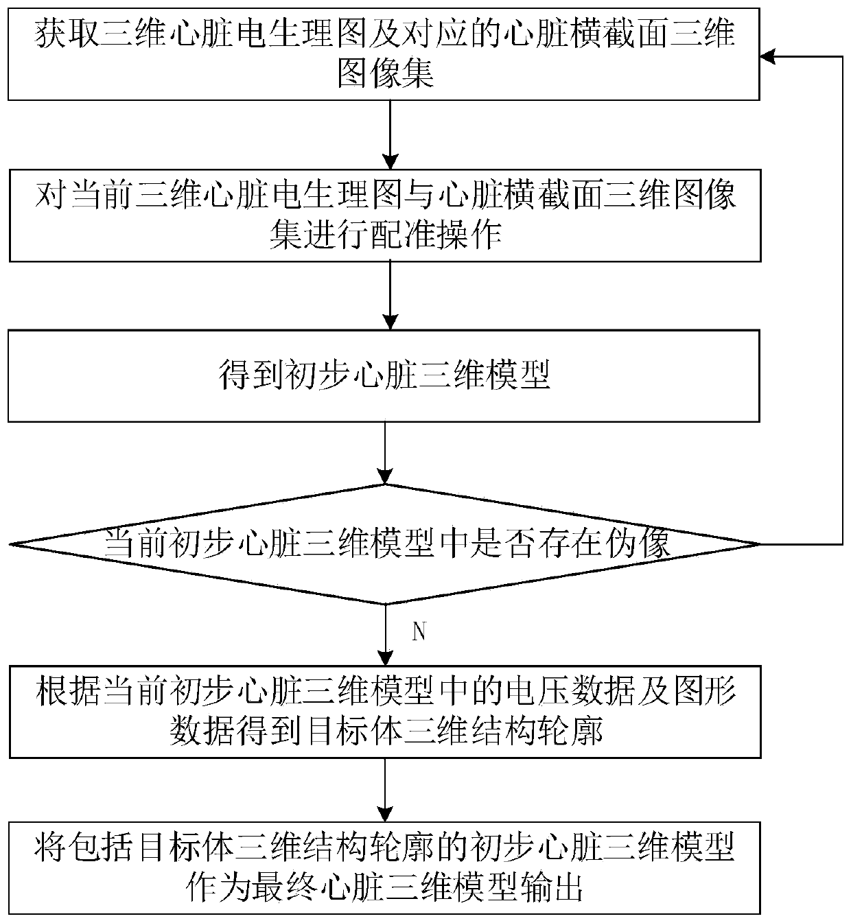
[0047] Such as figure 1 As shown, this embodiment provides a method for constructing a three-dimensional model of the heart used in cardiac radiotherapy, including the following steps:

[0048] Obtain a three-dimensional cardiac electrophysiological map and a corresponding three-dimensional image set of cardiac cross-section;

[0049] Perform a registration operation on the current 3D cardiac electrophysiological map and the 3D cross-sectional image set of the heart to obtain a preliminary 3D model of the heart;

[0050] Judging whether there are artifacts in the current preliminary three-dimensional heart model, if yes, then re-acquire the three-dimensional cardiac electrophysiological map and / or three-dimensional image set of cardiac cross-section, and then perform the registration operation again, if not, then according to the current preliminary three-dimensional cardiac model The electrophysiological data and graphic data are used to obtain the three-dimensional structur...

Embodiment 2

[0056] The technical solution provided in this embodiment is a further improvement made on the basis of the technical solution in embodiment 1. The difference between this embodiment and embodiment 1 is:

[0057] In this embodiment, when obtaining the preliminary three-dimensional model of the heart, the specific steps are as follows:

[0058] Process the data format of the current three-dimensional cardiac electrophysiological map into DICOM RT format to obtain the process three-dimensional cardiac electrophysiological map;

[0059] Acquiring multiple cardiac cross-sectional images in the current cardiac cross-sectional three-dimensional image set;

[0060] Using the registration method based on point-line-surface geometric feature constraints, the process 3D cardiac electrophysiological map and multiple cardiac cross-sectional images are processed to obtain a preliminary 3D model of the heart. In the registration operation, the 3D coordinates of the process 3D cardiac electr...

Embodiment 3

[0063] The technical solution provided in this embodiment is a further improvement made on the basis of the technical solution in embodiment 1 or 2. The difference between this embodiment and embodiment 1 or 2 is:

[0064] In this embodiment, an artifact detection algorithm based on deep learning is used to determine whether there are artifacts in the current preliminary three-dimensional model of the heart.


PUM


Abstract

The invention belongs to the technical field of model construction, and discloses a heart three-dimensional model construction method for cardiac radiotherapy. The method comprises the following steps: acquiring a three-dimensional heart electrophysiology map and a corresponding heart cross section three-dimensional image set; performing registration operation to obtain a preliminary heart three-dimensional model; judging whether an artifact exists in the current preliminary heart three-dimensional model or not; if yes, repeating the above steps, and if not, obtaining a target body three-dimensional structure contour; and outputting the preliminary heart three-dimensional model comprising the three-dimensional structure contour of the target body as a final heart three-dimensional model. According to the invention, the final heart three-dimensional model which can be used for heart radiotherapy and has reference significance is generated through the three-dimensional heart electrophysiology map and the heart cross section three-dimensional image set; the ventricular tachycardia can be treated on the basis of the ray ablation target point of the standard radiosurgical equipment, sothat the treatment time of the ventricular tachycardia is effectively shortened, the treatment process can be simplified to a certain extent, and the ventricular tachycardia treatment device is suitable for popularization and application.

Description

technical field [0001] The invention belongs to the technical field of model construction, and in particular relates to a method for constructing a three-dimensional heart model used in cardiac radiotherapy. Background technique [0002] Ventricular tachycardia is an irregular heartbeat of the heart. The current conventional treatment for ventricular tachycardia is to insert a catheter into the heart. The catheter then records voltage data from a large number of points on the inner surface of the heart to form a voltage map. The doctor then analyzes the voltage. map, and define the target area for ablation, and then send blocking voltage through the catheter in the target area, and then bring thermal energy to the target location to change the rhythmic activation cycle of the heart. Catheter treatment of ventricular tachycardia as described above is usually effective, but for the case where the target area is located inside the heart, catheter therapy will cause more damage ...

Claims


Application Information

Patent Timeline
Patent Type & Authority Applications(China)
IPC IPC(8): G06T17/00G16H30/20G16H50/50
CPCG06T17/00G16H30/20G16H50/50G06T2210/41
Inventor 韦崇高
Owner 韦崇高
Learn More